Review
. 2011 Mar 23:4:41.
doi: 10.1186/1756-3305-4-41.

Zoonotic helminths affecting the human eye

Affiliations
Review

Zoonotic helminths affecting the human eye

Domenico Otranto et al. Parasit Vectors. .

Abstract

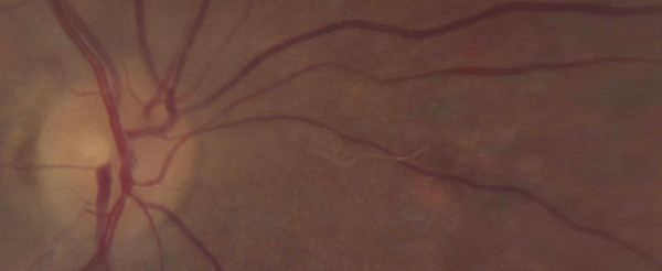
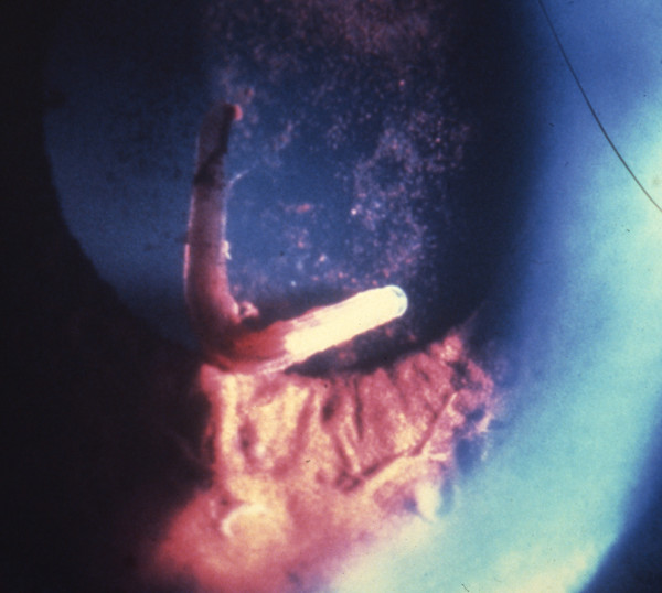
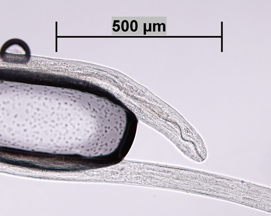
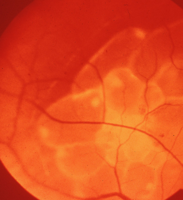
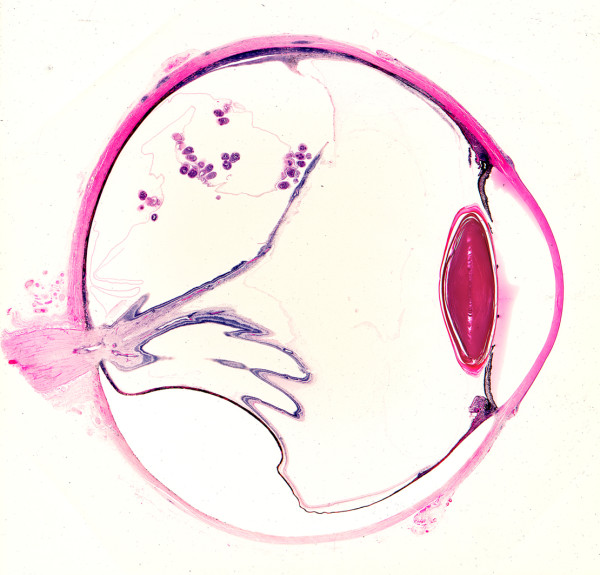
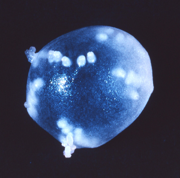
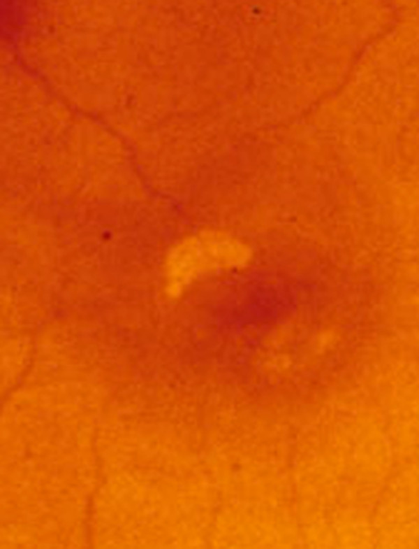
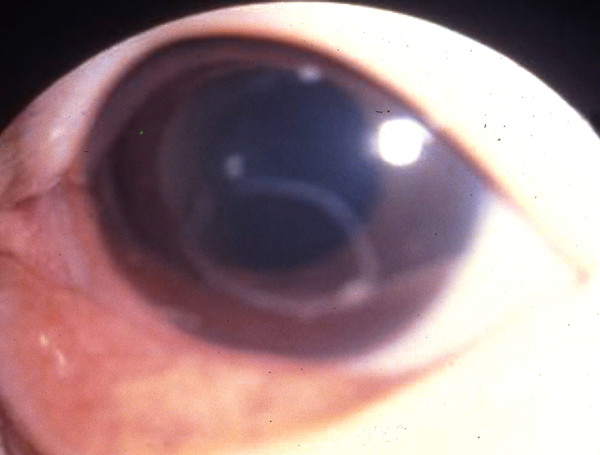
Nowaday, zoonoses are an important cause of human parasitic diseases worldwide and a major threat to the socio-economic development, mainly in developing countries. Importantly, zoonotic helminths that affect human eyes (HIE) may cause blindness with severe socio-economic consequences to human communities. These infections include nematodes, cestodes and trematodes, which may be transmitted by vectors (dirofilariasis, onchocerciasis, thelaziasis), food consumption (sparganosis, trichinellosis) and those acquired indirectly from the environment (ascariasis, echinococcosis, fascioliasis). Adult and/or larval stages of HIE may localize into human ocular tissues externally (i.e., lachrymal glands, eyelids, conjunctival sacs) or into the ocular globe (i.e., intravitreous retina, anterior and or posterior chamber) causing symptoms due to the parasitic localization in the eyes or to the immune reaction they elicit in the host. Unfortunately, data on HIE are scant and mostly limited to case reports from different countries. The biology and epidemiology of the most frequently reported HIE are discussed as well as clinical description of the diseases, diagnostic considerations and video clips on their presentation and surgical treatment.


Figures

Figure 1
Figure 1
Nematode larva in the retinal fundus. Larva, presumably of Baylisascaris procyonis, in the retinal fundus of a patient. The infection was presumed to have been acquired in Connecticut, USA. Larva measures approximately 1.4 mm and was treated with retinal lasar. (Original; courtesy of Drs. Caplivski, Bhatnagar, and Goldberg, Mount Sinai School of Medicine).
Figure 2
Figure 2
Gnathostoma spinigerum larva in the anterior chamber of the eye. Gnathostoma spinigerum larva in the anterior chamber of the eye of patient in Thailand. (From Teekhasaenee C, Richt R, Kanchanaranya. Ocular parasitic infection in Thailand. Rev Inf Dis. 1986, 8:350-356).
Figure 3
Figure 3
Acanthocheilonema - Mansonella -like worm. Anterior end of an Acanthocheilonema - Mansonella -like worm removed from the posterior chamber of the vitreous of a patient in Kansas, USA. Scale bar = 500 μm. Original; courtesy of DPDx, CDC.
Figure 4
Figure 4
Cysticercus cyst of Taenia crassiceps in situ. Retinal photograph showing budding cysticercus of Taenia crassiceps in situ. (From the collection of Herman Zaiman, "A Presentation of Pictorial Parasites").
Figure 5
Figure 5
Coenurus cyst behind displaced retina. Sagittal section of eye from Ghanaian showing coenurus cyst with multiple protoscoleces lying behind displaced retina. (From Parasites In Human Tissues, Orihel and Ash, ASCP Press, 1995).
Figure 6
Figure 6
Coenurus cyst after surgical removing from the eye. Intact coenurus cyst removed from subconjunctival tissue an Ugandan child showing multiple protoscoleces. (Original by Paul Beaver).
Figure 7
Figure 7
Alaria sp. in the eye. Freely moving Alaria mesocercaria on the retina of the eye. The anterior sucker is evident on the left side of the organism. (From the collection of Herman Zaiman, "A Presentation of Pictorial Parasites").
Figure 8
Figure 8
Angiostrongylus cantonensis in the anterior chamber of the eye. Angiostrongylus cantonensis in the anterior chamber. (Original by John Cross, courtesy of Lawrence Ash).
Figure 9
Figure 9
Thelazia calllipaeda in a heavily infected dog. Heavy infection by Thelazia callipaeda nematodes in the conjunctiva of a dog from Italy.
Figure 10
Figure 10
Thelazia californiensis from a human patient. Posterior end of a female Thelazia californiensis from the conjunctiva of a human patient in New Hampshire, USA showing cuticle serration. Scale bar = 50 μm. Original; courtesy of DPDx, CDC.
Figure 11
Figure 11
Zoonotic Onchocerca from human ocular connective tissue. Zoonotic Onchocerca sp. from a nodular granuloma of the eye in a patient from Ohio, USA. a) Transverse section of female worm shown in Fig. 5a encased in a nodular granuloma. Low cuticular ridges and inner striae, 2 per ridge, are evident Hematoxylin and eosin stain. Scale bar = 50 μm. (Original; courtesy of Drs. Yassin and Hariri, University of Pittsburg Medical Center). b) Short piece of female Onchocerca sp. removed from granuloma tissue before fixation, showing characteristic, diagnostic structures of the cuticle with circular ridges and inner cuticular striae. Scale bar = 150 μm. (Original; courtesy of Drs. Yassin and Hariri, University of Pittsburg Medical Center).

Similar articles

Cited by

References

    1. Williams RA, Brody BL, Thomas RG, Kaplan RM, Brown SI. The psychosocial impact of macular degeneration. Arch Ophthalmol. 1998;116:514–520. - PubMed
    1. Blindness and poverty. http://webadmin.hollows.org/Assets/Files/Blindness%20and%20Poverty%5B1%5...
    1. World Health Organization 2003. Onchocerciasis. http://www.who.int/blindness/partnerships/onchocerciasis_home/en/index.html
    1. Ryan E, Durrand M. In: Tropical Infectious Diseases Principals, Pathogens, and Practices. 2. Guerrant RL, WalkerDH, Weller PF, editor. Philadelphia: Churchill Livingstone; 2005. Ocular Disease; pp. 1554–1600.
    1. Cutler SJ, Fooks AR, van der Poel WH. Public health threat of new, reemerging, and neglected zoonoses in the industrialized world. Emerg Infect Dis. 2010;16(1):1–7. doi: 10.3201/eid1601.081467. - DOI - PMC - PubMed

Publication types

MeSH terms