. 2011 Mar 23:12:22.
doi: 10.1186/1471-2172-12-22.

Human CD8 T cells generated in vitro from hematopoietic stem cells are functionally mature

Affiliations

Human CD8 T cells generated in vitro from hematopoietic stem cells are functionally mature

Génève Awong et al. BMC Immunol. .

Abstract

Background: T cell development occurs within the highly specialized thymus. Cytotoxic CD8 T cells are critical in adaptive immunity by targeting virally infected or tumor cells. In this study, we addressed whether functional CD8 T cells can be generated fully in vitro using human umbilical cord blood (UCB) hematopoietic stem cells (HSCs) in coculture with OP9-DL1 cells.

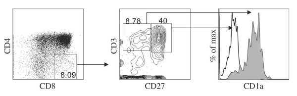
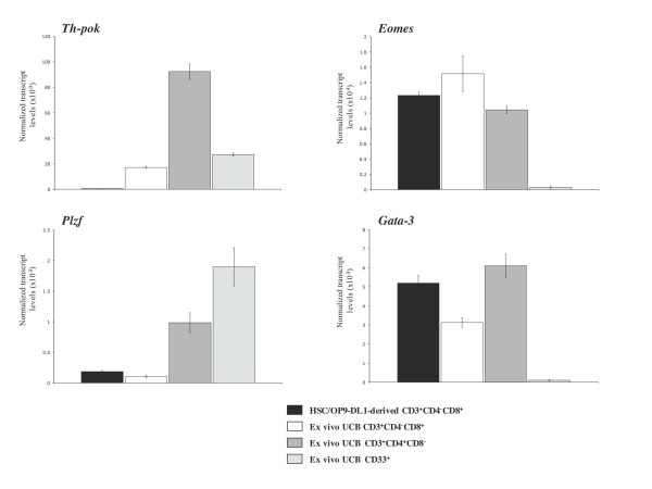
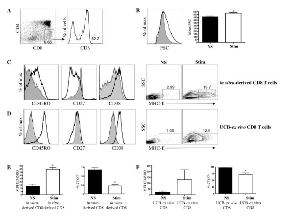
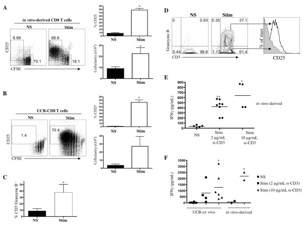
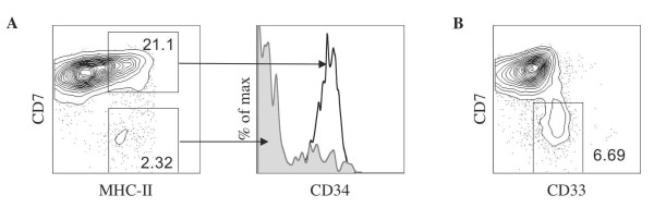
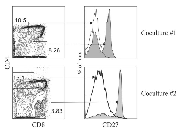
Results: HSC/OP9-DL1 cocultures supported the differentiation of CD8 T cells, which were TCR/CD3(hi) CD27(hi) CD1a(neg) and thus phenotypically resembled mature functional CD8 single positive thymocytes. These in vitro-generated T cells also appeared to be conventional CD8 cells, as they expressed high levels of Eomes and low levels of Plzf, albeit not identical to ex vivo UCB CD8 T cells. Consistent with the phenotypic and molecular characterization, upon TCR-stimulation, in vitro-generated CD8 T cells proliferated, expressed activation markers (MHC-II, CD25, CD38), secreted IFN-γ and expressed Granzyme B, a cytotoxic T-cell effector molecule.

Conclusion: Taken together, the ability to direct human hematopoietic stem cell or T-progenitor cells towards a mature functional phenotype raises the possibility of establishing cell-based treatments for T-immunodeficiencies by rapidly restoring CD8 effector function, thereby mitigating the risks associated with opportunistic infections.


Figures

Figure 1
Figure 1
Characterization of CD8+ T cells generated in vitro. Flow cytometric analysis for the expression of CD8 and CD4 from human UCB-derived HSCs cultured on OP9-DL1 cells for 65 days. CD8+ CD4- single positive (SP) cells were gated, as indicated, and analyzed for the expression of CD27 and CD3, with CD1a expression shown for cells gated as CD3+ CD27- or CD27+ (shaded and clear histograms, respectively). Data are representative of over 6 experiments for CD8/CD4 expression and of 3 experiments for CD27/CD1a expression.
Figure 2
Figure 2
Gene expression analysis of CD8+ SP T cells obtained from HSCs cultured on OP9-DL1 cells. Gene expression analysis by QRT-PCR from coculture-derived CD8 SP T cells (CD8+ CD4- CD3+). CD8 SP, CD4 SP T cells and CD33+ myeloid cells were purified from the Lin+ fraction of UCB samples and served as controls. Transcript levels for the indicated genes were normalized to human β-actin, and these data are representative of 3 independent experiments, with the standard error bars shown corresponding to values obtained from triplicate wells within an individual experiment.
Figure 3
Figure 3
Characterization of activation marker status on activated CD8+ T cells generated in vitro. (A) Day 60-70 HSC/OP9-DL1 coculture-derived CD8 SP T cells were purified as shown, CD8+ CD4- and CD3+, and stimulated anti-CD3/CD28 mAbs for 5 days. (B) Left-side panel displays flow cytometric analyses of cell size measured by Forward-light Scatter (FSC) intensity; right-side panel displays mean FSC for non-stimulated (NS) and stimulated (Stim) CD8+ CD3+ cells (n = 5; p = 0.004; t-test). (C) Flow cytometric analyses for the expression of CD45RO, CD27, CD38 and MHC-class II for stimulated (Stim) and non-stimulated (NS) CD8+ CD3+ cells (solid black line and shaded histograms, respectively). (D) Flow cytometric analyses for the expression of CD45RO, CD27, CD38 and MHC-class II for stimulated (Stim) and non-stimulated (NS) CD8+ CD3+ cells obtained from the UCB Lin+ fraction. Analysis of CD45RO mean fluorescence intensity (MFI) and percentage of CD27+ cells for (E) HSC/OP9-DL1 coculture (in-vitro)-derived CD8+ CD3+ T cells and (F) UCB-ex vivo CD8+ CD3+ T cells were performed (MFI CD45RO in vitro-derived, p = 0.02; n = 3; t-test; %CD27+ in vitro-derived, p = 0.007; n = 3; t-test; %CD27+ UCB-ex vivo, p = 0.03; n = 2; t-test). * indicates statistical significance.
Figure 4
Figure 4
Functional characterization on activated CD8+ T cells generated in vitro. (A) Day 60-70 HSC/OP9-DL1 coculture-derived CD8 SP T cells and (B) UCB-ex vivo CD8 T cells were purified and stimulated with anti-CD3/CD28 mAbs for 5 days. Flow cytometric analyses of CFSE levels and CD25 (left side), and percentage of CD25+ cells (upper right) and cellularity (lower right) for stimulated (Stim) versus non-stimulated (NS) are shown (* indicates p < 0.05). (C) Analysis of the percentage of in vitro-derived CD8+ CD3+ intracellular Granzyme-B+ expressing cells for stimulated versus non-stimulated is shown (p = 0.02). (E-F) Human IFNγ levels from culture supernatants derived from the above-outlined experiment were determined by ELISA. (E) Statistical significance was measured by unpaired t-test for stimulated and non-stimulated in vitro-derived T cells. * (p < 0.005) 2 μg/mL anti-CD3/CD28 stimulated group versus non-stimulated. ** (p < 0.0005) 10 μg/mL anti-CD3/CD28 versus non-stimulated. Data are representative of at least 3 independent experiments, with the exception of the data from the 10 μg/ml stimulations, which are derived from 2 independent experiments. (F) Statistical significance was measured by unpaired t-test for stimulated (10 μg/mL) and non-stimulated UCB-ex vivo T cells. * (p < 0.05). Data are representative of at least 4 independent UCB-ex vivo CD8 T cells. The data from the in vitro-generated T cells (non-stimulated and 10 μg/mL stimulation), are derived from 2 independent experiments.
Figure 5
Figure 5
Characterization of MHC class II-expressing cells derived from OP9-DL1 culture. Flow cytometric analysis for cell surface expression of (A) MHC class II, CD7 (left side) and CD34 on MHC II+ CD7+-gated cells (solid black line) and MHC II+ CD7--gated cells (shaded histogram) from a day 40 HSC/OP9-DL1 coculture, and (B) CD33 and CD7 from a day 45 HSC/OP9-DL1 coculture. Numbers in plots indicate percentage of cells within each gate shown. Data are representative of at least 2 independent experiments.
Figure 6
Figure 6
Characterization of single-positive T cells generated from HSC/OP9-DL1 coculture. Flow cytometric analysis for cell surface expression of CD4 and CD8 is presented for 2 independent cocultures (left side). CD27 expression was examined on CD4+ CD8--gated cells (solid black line) and on CD4- CD8+-gated cells (shaded histogram).

References

    1. Spits H. Development of alphabeta T cells in the human thymus. Nat Rev Immunol. 2002;2:760–772. doi: 10.1038/nri913. - DOI - PubMed
    1. Storek J, Gooley T, Witherspoon RP, Sullivan KM, Storb R. Infectious morbidity in long-term survivors of allogeneic marrow transplantation is associated with low CD4 T cell counts. Am J Hematol. 1997;54:131–138. doi: 10.1002/(SICI)1096-8652(199702)54:2<131::AID-AJH6>3.0.CO;2-Y. - DOI - PubMed
    1. Douek DC, Vescio RA, Betts MR, Brenchley JM, Hill BJ, Zhang L, Berenson JR, Collins RH, Koup RA. Assessment of thymic output in adults after haematopoietic stem-cell transplantation and prediction of T-cell reconstitution. Lancet. 2000;355:1875–1881. doi: 10.1016/S0140-6736(00)02293-5. - DOI - PubMed
    1. van den Brink MR, Alpdogan O, Boyd RL. Strategies to enhance T-cell reconstitution in immunocompromised patients. Nat Rev Immunol. 2004;4:856–867. doi: 10.1038/nri1484. - DOI - PubMed
    1. Schmitt TM, Zúñiga-Pflücker JC. Induction of T cell development from hematopoietic progenitor cells by delta-like-1 in vitro. Immunity. 2002;17:749–756. doi: 10.1016/S1074-7613(02)00474-0. - DOI - PubMed

Publication types

MeSH terms