. 2011 May 10;413(2):253-64.
doi: 10.1016/j.virol.2011.02.022. Epub 2011 Mar 22.

N-linked glycosylation of dengue virus NS1 protein modulates secretion, cell-surface expression, hexamer stability, and interactions with human complement

Affiliations

N-linked glycosylation of dengue virus NS1 protein modulates secretion, cell-surface expression, hexamer stability, and interactions with human complement

Pawit Somnuke et al. Virology. .

Abstract

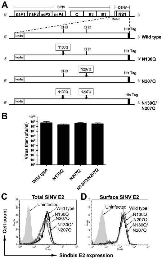
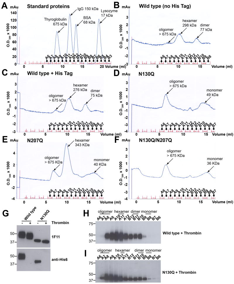
Dengue virus (DENV) NS1 is a versatile non-structural glycoprotein that is secreted as a hexamer, binds to the cell surface of infected and uninfected cells, and has immune evasive functions. DENV NS1 displays two conserved N-linked glycans at N130 and N207. In this study, we examined the role of these two N-linked glycans on NS1 secretion, stability, and function. Because some groups have reported reduced yields of infectious DENV when N130 and N207 are changed, we analyzed glycosylation-deficient NS1 phenotypes using a transgenic expression system. We show that the N-linked glycan at position 130 is required for stabilization of the secreted hexamer whereas the N-linked glycan at residue 207 facilitates secretion and extracellular protein stability. Moreover, NS1 mutants lacking an N-linked glycan at N130 did not interact efficiently with complement components C1s and C4. In summary, our results elucidate the contribution of N-linked glycosylation to the function of DENV NS1.


Figures

Figure 1
Figure 1. Expression of wild type and N-linked glycan mutant DENV NS1 does not differentially affect SINV replication
(A) Schematic diagram of SINV vector encoding DENV NS1 constructs. Wild type or mutants with a glutamine substitution at the first (N130Q), second (N207Q), or both glycosylation sites (N130Q/N207Q) of DENV-2 NS1 were inserted into SINV 39HK vector backbone after the nonstructural nsP1-4 and structural proteins, capsid, envelope 1 and 2. The leader sequence was derived from the last 24 amino acids of DENV-2 E protein. A hexa-histidine tag (His6 Tag) was added onto the carboxy-terminus of each construct. The boxes indicate glutamine for asparagine substitutions within the glycosylation sites and arrows indicate the sites of mutations. CHO indicates the position of N-linked glycans. (B-D) Transgenic expression of variant DENV NS1 does not affect SINV replication. BHK21 cells were infected with SINV expressing wild type and NS1 mutants at an MOI of 1.0. The supernatants were collected 18 h post-infection and subjected to plaque assay (B). Level of SINV infection was also assessed by total (C) and surface (D) SINV-E2 staining with mouse hyper-immune serum against SINV.
Figure 2
Figure 2. N-linked glycan (N207) is important for surface localization and secretion of DENV NS1
(A-E) Decreased surface localization and secretion was observed in N207Q and N130Q/N207Q mutant NS1. Infected cells were harvested at 18 h post-infection and total (A) and surface (B) DENV NS1 expression was determined by immunofluorescence using 1F11 anti-DENV-2 NS1 MAb, which recognizes a linear epitope on NS1. Levels of secreted NS1 were quantified by ELISA of supernatants harvested at 18 h post-infection (C, open bars). To deplete cellular pools of sulfated GAGs, cells were treated with 20 mM sodium chlorate at 6 h after infection and the cells and supernatants were harvested 12 h later for analysis of total (D) and surface NS1 (E) by immunofluorescence using NS1-specific MAb 1F11 and secreted NS1 levels by ELISA (C, filled bars) respectively. (F-I) Conformation-dependent anti-NS1 antibodies did not efficiently recognize N207 mutants. Infected cells were harvested at 18 h post-infection and total (F and H) and surface (G and I) DENV NS1 expression was determined using the conformational-dependent cross-reactive 9NS1 MAb (F and G) and polyclonal anti-DENV-2 NS1 (H and I). J. Immunoreactivity of purified wild type and NS1 mutants. Equal amounts (100 ng) of wild type and mutant NS1 were boiled prior to 4-12% SDS-PAGE in the presence (reducing) or absence (non-reducing) of β-mercaptoethanol. Western blot analysis was performed subsequently with 1F11, 9NS1 or polyclonal anti-NS1. Error bars indicate standard error of the mean (SEM) from five independent experiments. Asterisks indicate statistical significant of NS1 secretion compared to wild type DENV2 NS1 (n.s., not significant; *, P < 0.05; ***, P < 0.0001). Histograms represent 3 to 5 independent experiments.
Figure 3
Figure 3. N-linked glycosylation does not affect secreted DENV NS1 from binding back to the cell surface
Wild type and mutant NS1 were purified by immunoaffinity chromatography. Serum-free supernatants from SINV-DENV-2-NS1 infected BHK21 cells at an MOI of 1.0 were collected at 18 to 20 h post-infection and purified over an anti-NS1 MAb 2G6 column. Samples of eluted fractions were boiled prior to 4-12% SDS-PAGE under non-reducing condition and analyzed by Western blot with 1F11 anti-DENV-2 NS1 MAb (wild type (A), N130Q (C), N207Q (E) and N130Q/N207Q (G)) and silver staining (wild type (B), N130Q (D), N207Q (F) and N130Q/N207Q (H)); the numbers below each gel correspond to the eluted fractions. (I-K) Binding of wild type and mutant NS1 to the cell surface depends on GAGs. Immunoaffinity purified NS1 (10 μg/ml) (I) or serum-free supernatants of SINV-DENV-2-NS1-infected cells containing 2 μg/ml of NS1 (J) were incubated with BHK21 cells and NS1 binding was detected by 2G6 anti-DENV-2 NS1 MAb. Binding was also performed after BHK21 cells were pre-treated with 20 mM sodium chlorate overnight and incubated with purified NS1 (K). Histograms are representative of 3 independent experiments.
Figure 4
Figure 4. N-linked glycan at N207 influences the rate of accumulation and stability of secreted DENV NS1
Secreted NS1 from SINV-DENV2-NS1 infected BHK cells (2 μg/ml) was treated with Endo H (A) or PNGase F (B) for 3 h at 37°C. Western blot analysis of enzyme digested NS1 was assessed by 4-12% SDS-PAGE under reducing condition using 1F11 anti-DENV2-NS1 MAb. (C and D) BHK21 cells infected at an MOI of one were harvested at 14 h and then were radiolabeled with [35S] and chased at different time points. Supernatants (C) and lysates (D) were collected, pre-cleared with isotype control Sepharose, and immunoprecipitated with 1F11 anti-DENV-2 NS1 MAb and protein A-Sepharose. (E-H) Western blot analysis of secreted NS1. Supernatants containing 2 μg/ml of wild type (E), N130Q (F), N207Q (G), and N130Q/N207Q (H) NS1 were incubated at 37°C prior to analysis by 12% SDS-PAGE under reducing conditions followed by western blotting with 1F11 anti-DENV2 NS1 MAb. Results are representative of two or three independent experiments. I. Using quantitative densitometry, the intensity of wild type, N130Q, N207Q, and N130Q/N207Q NS1 at each time point was compared to level of NS1 (100%) at the starting time (0 h). Error bars indicate standard error of the mean corresponding to three independent experiments.
Figure 5
Figure 5. The N-linked glycan at position N130 is required for stabilization of the secreted hexamer
(A-F) Size exclusion chromatography of immunoaffinity purified DENV-NS1. The peak fractions of NS1 were pooled and adjusted to 30 μg/ml. Size exclusion profiles of affinity-purified NS1 was determined by injecting 1 ml of sample onto a Superdex 200 column. Samples included standard proteins (A), wild type NS1 without hexa-histidine tag (B), wild type NS1 with hexa-histidine tag (C), N130Q (D), N207Q (E) and N130Q/N207Q (F). (G-I) Size exclusion chromatography of NS1 lacking hexa-histidine tag in bulk supernatants. Serum-free supernatants harvested from SINV-DENV-2-wild type or N130Q NS1-infected BHK cells at 18 h post-infection were treated with 0.05 unit of thrombin for 16 h at room temperature and concentrated 50-fold. A portion of thrombin-treated NS1 was subjected to 4-10% SDS-PAGE under reducing condition, followed by Western blotting with 1F11 anti-NS1 MAb or anti-His6 antibody (G). Thrombin-treated wild type or N130Q supernatants (1 ml) were injected onto a Superdex 200 column. Samples of eluted fractions were boiled prior to 4-12% SDS-PAGE under reducing condition and analyzed by Western blot with 1F11 anti-DENV-2 NS1 (Wild type (H), N130Q (I)). Results are representative of three to four independent experiments.
Figure 6
Figure 6. Loss of N-linked glycan at position N130 affects NS1 binding to human complement components
Equal amounts (5 μg/ml) of NS1 in culture supernatants were used in complement binding ELISA. Concentration was determined by: (A) direct coating of NS1 to microtiter plates and detection with anti-His6 tag MAb, and (B) Western blot with 1F11 anti-DENV-2 NS1. Binding of wild type and mutant NS1 to human complement components was assessed by ELISA (C). Microtiter plates were coated with BSA, complement proteins C4, C4b, C1s proenzyme or C1s (15 μg/ml). Supernatants with equal concentrations (5 μg/ml) of wild type or mutant NS1 from SINV-DENV-2-NS1-infected cells were added and bound NS1 was detected. (D) Hexameric NS1 interacts most efficiently with complement components. Immunoaffinity purified wild type NS1 hexamer and dimer isolated from a Superdex 200 column at equimolar concentration (40 nM) were added to BSA or complement components and bound NS1 was detected with anti-His6 tag MAb. Error bars indicate standard error of the mean corresponding to 3 or 4 independent experiments. Asterisks indicate binding that is statistically different compared to (C) wild type DENV2 NS1 or (D) hexameric NS1 (n.s., not significant; *, P < 0.05).

References

    1. Alcon-LePoder S, Drouet MT, Roux P, Frenkiel MP, Arborio M, Durand-Schneider AM, Maurice M, Le Blanc I, Gruenberg J, Flamand M. The secreted form of dengue virus nonstructural protein NS1 is endocytosed by hepatocytes and accumulates in late endosomes: implications for viral infectivity. J Virol. 2005;79(17):11403–11. - PMC - PubMed
    1. Alcon S, Talarmin A, Debruyne M, Falconar A, Deubel V, Flamand M. Enzyme-linked immunosorbent assay specific to Dengue virus type 1 nonstructural protein NS1 reveals circulation of the antigen in the blood during the acute phase of disease in patients experiencing primary or secondary infections. J Clin Microbiol. 2002;40(2):376–81. - PMC - PubMed
    1. Avirutnan P, Fuchs A, Hauhart RE, Somnuke P, Youn S, Diamond MS, Atkinson JP. Antagonism of the complement component C4 by flavivirus nonstructural protein NS1. J Exp Med. 2010;207(4):793–806. - PMC - PubMed
    1. Avirutnan P, Punyadee N, Noisakran S, Komoltri C, Thiemmeca S, Auethavornanan K, Jairungsri A, Kanlaya R, Tangthawornchaikul N, Puttikhunt C, Pattanakitsakul SN, Yenchitsomanus PT, Mongkolsapaya J, Kasinrerk W, Sittisombut N, Husmann M, Blettner M, Vasanawathana S, Bhakdi S, Malasit P. Vascular leakage in severe dengue virus infections: a potential role for the nonstructural viral protein NS1 and complement. J Infect Dis. 2006;193(8):1078–88. - PubMed
    1. Avirutnan P, Zhang L, Punyadee N, Manuyakorn A, Puttikhunt C, Kasinrerk W, Malasit P, Atkinson JP, Diamond MS. Secreted NS1 of dengue virus attaches to the surface of cells via interactions with heparan sulfate and chondroitin sulfate E. PLoS Pathog. 2007;3(11):e183. - PMC - PubMed

Publication types

Substances