. 2011 Jun;209(3):327-35.
doi: 10.1530/JOE-10-0493. Epub 2011 Mar 23.

The effects of ACTH on steroid metabolomic profiles in human adrenal cells

Affiliations

The effects of ACTH on steroid metabolomic profiles in human adrenal cells

Yewei Xing et al. J Endocrinol. 2011 Jun.

Abstract

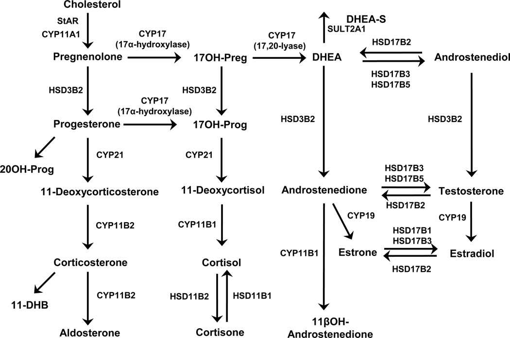
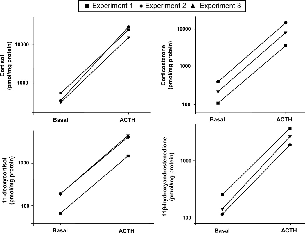
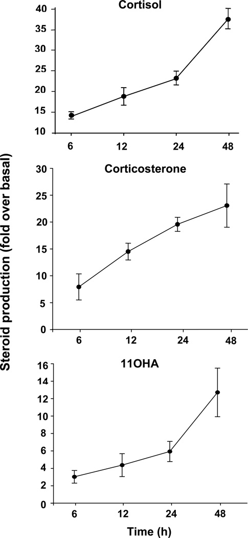
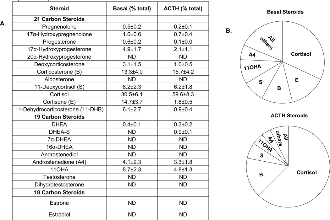
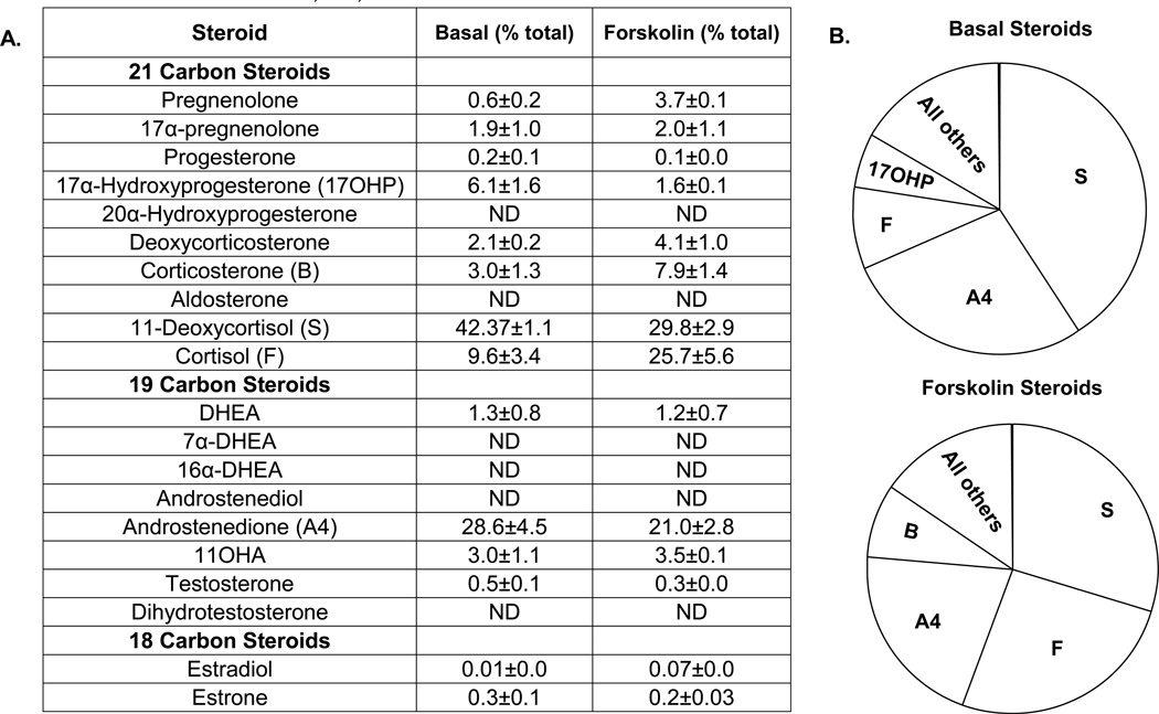
The adrenal glands are the primary source of mineralocorticoids, glucocorticoids, and the so-called adrenal androgens. Under physiological conditions, cortisol and adrenal androgen synthesis are controlled primarily by ACTH. Although it is well established that ACTH can stimulate steroidogenesis in the human adrenal gland, the effect of ACTH on overall production of different classes of steroid hormones has not been defined. In this study, we examined the effect of ACTH on the production of 23 steroid hormones in adult adrenal primary cultures and 20 steroids in the adrenal cell line, H295R. Liquid chromatography/tandem mass spectrometry analysis revealed that, in primary adrenal cell cultures, cortisol and corticosterone were the two most abundant steroid hormones produced with or without ACTH treatment (48 h). Cortisol production responded the most to ACTH treatment, with a 64-fold increase. Interestingly, the production of two androgens, androstenedione and 11β-hydroxyandrostenedione (11OHA), that were also produced in large amounts under basal conditions significantly increased after ACTH incubation. In H295R cells, 11-deoxycortisol and androstenedione were the major products under basal conditions. Treatment with forskolin increased the percentage of 11β-hydroxylated products, including cortisol and 11OHA. This study illustrates that adrenal cells respond to ACTH through the secretion of a variety of steroid hormones, thus supporting the role of adrenal cells as a source of both corticosteroids and androgens.


Figures

Fig. 1
Fig. 1. Steroid biosynthesis pathways
Summary of the steroidogenic pathways leading to synthesis of glucocorticoids, mineralocorticoids, androgens and estrogens.
Fig. 2
Fig. 2. Major steroids produced by AA cells with/without ACTH treatment
Primary human adrenal culture cells were isolated as described in Materials and Methods, and plated at a density of 300,000 cells/well in 24-well dishes. Cells were treated with/without ACTH (10 nM) for 48 h before harvest. The concentration of steroids in the medium was measured by LC-MS/MS and normalized to the amount of protein. The amount of four major steroids produced by AA cells, cortisol, corticosterone, 11-deoxycortisol and 11OHA are shown in the graphs with each line representing an individual, independent experiment.
Fig. 3
Fig. 3. Time-dependent stimulation of three major steroids produced in AA cells by ACTH treatment
Primary human adrenal culture cells were isolated as described in Materials and Methods, and plated at a density of 300,000 cells/well in 24-well dishes. Cells were treated with/without ACTH (10 nM) for indicated times. The concentration of steroids (cortisol, corticosterone and 11OHA) in the medium was measured by immunoassay and expressed as fold changes over basal level. Results represent means ± S.E.M of data from at least three experiments using cells isolated from different adrenal glands.
Figure 4
Figure 4. Percentage of major steroids produced by AA cells with/ without ACTH treatment
Primary human adrenal cells were isolated as described in Materials and Methods, and plated at a density of 300,000 cells/well in 24-well dishes. Cells were treated with/without ACTH (10nM) in 0.1% experimental medium for 48 h before harvest. The concentration of steroids in the medium was measured by LC-MS/MS and normalized to the amount of protein. Percentage of each steroid was calculated by dividing the amount of individual steroid with total steroid. A. Table summarizing the percentage of individual steroid compared to total steroid produced by AA cells. B. Pie chart summarizing the relative percentage of the major steroids produced in AA cells. Data represent results from three independent experiments and shown as means ± S.E.M; ND, not detectable.
Figure 5
Figure 5. Percentage of major steroids produced by H295R cells with/ without forskolin treatment
Three independent strains of the H295R adrenal cell model were cultured as described in Materials and Methods, and plated at a density of 400,000 cells/well in 12-well dishes. Cells were treated with/without forskolin (10 µM) in 0.1% experimental medium for 48 h before harvest. The concentration of steroids in the medium was measured by LC-MS/MS. Percentage of each steroid was calculated by dividing the amount of individual steroid with total steroid. A. Table summarizing the percentage of individual steroid compared to total steroid produced by H295R cells. B. Pie chart summarizing the relative percentage of four major steroids produced in H295R cells. Data represent results from three independent experiments and shown as means ± S.E.M; ND, not detectable.

Similar articles

Cited by

References

    1. Arvat E, Di Vito L, Lanfranco F, Maccario M, Baffoni C, Rossetto R, Aimaretti G, Camanni F, Ghigo E. Stimulatory effect of adrenocorticotropin on cortisol, aldosterone, and dehydroepiandrosterone secretion in normal humans: dose-response study. J Clin Endocrinol Metab. 2000;85:3141–3146. - PubMed
    1. Bassett MH, Suzuki T, Sasano H, De Vries CJ, Jimenez PT, Carr BR, Rainey WE. The orphan nuclear receptor NGFIB regulates transcription of 3beta-hydroxysteroid dehydrogenase. implications for the control of adrenal functional zonation. J Biol Chem. 2004;279:37622–37630. - PubMed
    1. Beishuizen A, Thijs LG, Vermes I. Decreased levels of dehydroepiandrosterone sulphate in severe critical illness: a sign of exhausted adrenal reserve? Crit Care. 2002;6:434–438. - PMC - PubMed
    1. Branchaud CL, Goodyer CG, Lipowski LS. Progesterone and estrogen production by placental monolayer cultures: effect of dehydroepiandrosterone and luteinizing hormone-releasing hormone. J Clin Endocrinol Metab. 1983;56:761–766. - PubMed
    1. Burger HG. Androgen production in women. Fertil Steril. 2002;77(Suppl 4):S3–S5. - PubMed

Publication types