Proximal tubular injury and rapid formation of atubular glomeruli in mice with unilateral ureteral obstruction: a new look at an old model
- PMID: 21429968
- PMCID: PMC3129891
- DOI: 10.1152/ajprenal.00022.2011
Proximal tubular injury and rapid formation of atubular glomeruli in mice with unilateral ureteral obstruction: a new look at an old model
Abstract
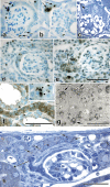
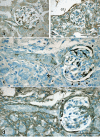
Unilateral ureteral obstruction (UUO), employed extensively as a model of progressive renal interstitial fibrosis, results in rapid parenchymal deterioration. Atubular glomeruli are formed in many renal disorders, but their identification has been limited by labor-intensive available techniques. The formation of atubular glomeruli was therefore investigated in adult male mice subjected to complete UUO under general anesthesia. In this species, the urinary pole of Bowman's capsule is normally lined by tall parietal epithelial cells similar to those of the proximal tubule, and both avidly bind Lotus tetragonolobus lectin. Following UUO, these cells became flattened, lost their affinity for Lotus lectin, and no longer generated superoxide (revealed by nitroblue tetrazolium infusion). Based on Lotus lectin staining, stereological measurements, and serial section analysis, over 80% of glomeruli underwent marked transformation after 14 days of UUO. The glomerulotubular junction became stenotic and atrophic due to cell death by apoptosis and autophagy, with concomitant remodeling of Bowman's capsule to form atubular glomeruli. In this degenerative process, transformed epithelial cells sealing the urinary pole expressed α-smooth muscle actin, vimentin, and nestin. Although atubular glomeruli remained perfused, renin immunostaining was markedly increased along afferent arterioles, and associated maculae densae disappeared. Numerous progressive kidney disorders, including diabetic nephropathy, are characterized by the formation of atubular glomeruli. The rapidity with which glomerulotubular junctions degenerate, coupled with Lotus lectin as a marker of glomerular integrity, points to new investigative uses for the model of murine UUO focusing on mechanisms of epithelial cell injury and remodeling in addition to fibrogenesis.
Figures


Similar articles
-
Fight-or-flight: murine unilateral ureteral obstruction causes extensive proximal tubular degeneration, collecting duct dilatation, and minimal fibrosis.Am J Physiol Renal Physiol. 2012 Jul 1;303(1):F120-9. doi: 10.1152/ajprenal.00110.2012. Epub 2012 Apr 25. Am J Physiol Renal Physiol. 2012. PMID: 22535799 Free PMC article.
-
Chronic unilateral ureteral obstruction in the neonatal mouse delays maturation of both kidneys and leads to late formation of atubular glomeruli.Am J Physiol Renal Physiol. 2013 Dec 15;305(12):F1736-46. doi: 10.1152/ajprenal.00152.2013. Epub 2013 Oct 9. Am J Physiol Renal Physiol. 2013. PMID: 24107422 Free PMC article.
-
Tubular obstruction leads to progressive proximal tubular injury and atubular glomeruli in polycystic kidney disease.Am J Pathol. 2014 Jul;184(7):1957-66. doi: 10.1016/j.ajpath.2014.03.007. Epub 2014 May 9. Am J Pathol. 2014. PMID: 24815352 Free PMC article.
-
Formation of atubular glomeruli in the developing kidney following chronic urinary tract obstruction.Pediatr Nephrol. 2011 Sep;26(9):1381-5. doi: 10.1007/s00467-010-1748-y. Epub 2011 Jan 9. Pediatr Nephrol. 2011. PMID: 21222000 Review.
-
Generation and evolution of atubular glomeruli in the progression of renal disorders.J Am Soc Nephrol. 2008 Feb;19(2):197-206. doi: 10.1681/ASN.2007080862. Epub 2008 Jan 16. J Am Soc Nephrol. 2008. PMID: 18199796 Review.
Cited by
-
The Fate of Nephrons in Congenital Obstructive Nephropathy: Adult Recovery is Limited by Nephron Number Despite Early Release of Obstruction.J Urol. 2015 Nov;194(5):1463-72. doi: 10.1016/j.juro.2015.04.078. Epub 2015 Apr 23. J Urol. 2015. PMID: 25912494 Free PMC article.
-
NADPH-oxidase 4 protects against kidney fibrosis during chronic renal injury.J Am Soc Nephrol. 2012 Dec;23(12):1967-76. doi: 10.1681/ASN.2012040373. Epub 2012 Oct 25. J Am Soc Nephrol. 2012. PMID: 23100220 Free PMC article.
-
ALTERATIONS IN HISTIDINE METABOLISM IS A FEATURE OF EARLY AUTOSOMAL DOMINANT POLYCYSTIC KIDNEY DISEASE (ADPKD).Trans Am Clin Climatol Assoc. 2024;134:47-65. Trans Am Clin Climatol Assoc. 2024. PMID: 39135565 Free PMC article.
-
Fight-or-flight: murine unilateral ureteral obstruction causes extensive proximal tubular degeneration, collecting duct dilatation, and minimal fibrosis.Am J Physiol Renal Physiol. 2012 Jul 1;303(1):F120-9. doi: 10.1152/ajprenal.00110.2012. Epub 2012 Apr 25. Am J Physiol Renal Physiol. 2012. PMID: 22535799 Free PMC article.
-
Regulated necrosis and failed repair in cisplatin-induced chronic kidney disease.Kidney Int. 2019 Apr;95(4):797-814. doi: 10.1016/j.kint.2018.11.042. Kidney Int. 2019. PMID: 30904067 Free PMC article.
References
Publication types
MeSH terms
Substances
Grants and funding
LinkOut - more resources
Full Text Sources
Other Literature Sources

