. 2011 May;138(9):1717-26.
doi: 10.1242/dev.059881. Epub 2011 Mar 23.

Arterial-venous network formation during brain vascularization involves hemodynamic regulation of chemokine signaling

Affiliations

Arterial-venous network formation during brain vascularization involves hemodynamic regulation of chemokine signaling

Jeroen Bussmann et al. Development. 2011 May.

Abstract

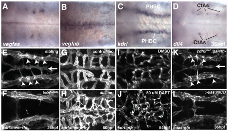
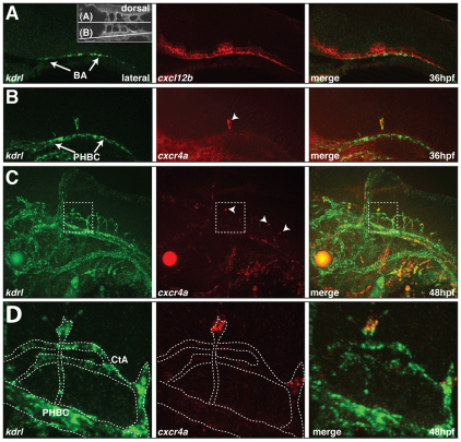
During angiogenic sprouting, newly forming blood vessels need to connect to the existing vasculature in order to establish a functional circulatory loop. Previous studies have implicated genetic pathways, such as VEGF and Notch signaling, in controlling angiogenesis. We show here that both pathways similarly act during vascularization of the zebrafish central nervous system. In addition, we find that chemokine signaling specifically controls arterial-venous network formation in the brain. Zebrafish mutants for the chemokine receptor cxcr4a or its ligand cxcl12b establish a decreased number of arterial-venous connections, leading to the formation of an unperfused and interconnected blood vessel network. We further find that expression of cxcr4a in newly forming brain capillaries is negatively regulated by blood flow. Accordingly, unperfused vessels continue to express cxcr4a, whereas connection of these vessels to the arterial circulation leads to rapid downregulation of cxcr4a expression and loss of angiogenic characteristics in endothelial cells, such as filopodia formation. Together, our findings indicate that hemodynamics, in addition to genetic pathways, influence vascular morphogenesis by regulating the expression of a proangiogenic factor that is necessary for the correct pathfinding of sprouting brain capillaries.


Figures

Fig. 1.
Fig. 1.
Development of the zebrafish hindbrain vasculature. (A) Maximal intensity projection of a confocal z-stack of a kdrl:gfp transgenic zebrafish brain at 60 hpf, indicating the position of the primordial hindbrain channels (PHBCs, blue), posterior communicating segments (PCSs, red), basilar artery (BA, red) and central arteries (CtAs, magenta). Dorsal view, anterior to the left. ov, otic vesicle; sc, spinal cord. Scale bar: 100 μm(B) Wire diagram of the hindbrain vasculature based on angiography at 60 hpf. Lateral oblique view. Color coding as in A. (C) Schematic representation of the hindbrain vascular network at 60 hpf in three individual wild-type (wt) embryos based on confocal z-stacks of kdrl:gfp transgenic embryos, indicating the position of PHBC-CtA connections (red filled circles), CtA-CtA connections (black dots) and CtA-BA/PCS connections (yellow filled circles). Dashed lines indicate connections without a visible lumen. Gray lines represent ventral (non-CtA) connections between the PHBC and the BA. (D) Schematic representation of stereotyped vascular connections in the hindbrain vascular network at 60 hpf. A and B, fixed CtA to BA connections; C, fixed PHBC to CtA connections. (E) Confocal time-lapse of BA formation in live kdrl:mem-rfp transgenic embryos between 30 and 40 hpf. Arrowheads indicate pruning of ventral non-CtA connections between the PHBC and the BA. (F-H) Confocal time-lapse of CtA formation in live kdrl:mem-rfp transgenic embryos. The boxed regions are enlarged in G and H, showing the presence of filopodial extensions on a non-lumenized CtA (arrowheads, G) but not on a lumenized CtA (arrow, H). (I) Schematic representation of BA and CtA formation. Transverse view. The dashed line indicates the planar level at which E and F were separated.
Fig. 2.
Fig. 2.
VegfA and Notch signaling are required for angiogenic sprouting in the zebrafish hindbrain. (A-D) Whole-mount in situ hybridization showing the expression of vegfaa (A), vegfab (B), kdrl (C) and dll4 (D) at 36 hpf. (E-L) Maximal intensity projections of confocal z-stacks at 36 (E,F,K,L), 54 (I,J) or 60 (G,H) hpf. Arrowheads indicate CtA sprouts emerging from the PHBC. Arrow indicates the position of the BA. (E,F) Sibling (E) or kdrl mutant (F) embryos, with endothelial cells labeled by kdrl:mem-rfp expression. (G,H) kdrl:mem-rfp-expressing embryos injected with control MO (G) or dll4 MO (H). (I,J) kdrl:gfp-expressing embryos treated from 30 to 54 hpf with DMSO (I) or DAPT (J). (K,L) Embryos from a cross between cdh5:gal4ff, uas:gfp and uas:NICD. The embryo in K is negative for uas:NICD, whereas that in L is heterozygous for uas:NICD. Endothelial cells are labeled by cdh5:gal4ff, uas:gfp expression. Dorsal views, anterior to the left.
Fig. 3.
Fig. 3.
Expression of cxcl12b and cxcr4a in the zebrafish hindbrain. (A-D) Two-color fluorescent in situ hybridization showing the distribution of kdrl mRNA (green) and cxcl12b (A) and cxcr4a (B-D) mRNA (red). Lateral views, anterior to the left. (A) cxcl12b and kdrl expression at 36 hpf. Inset indicates the plane of view in A and B. (B) cxcr4a and kdrl expression at 36 hpf. Arrowhead indicates a single cxcr4a-expressing CtA sprout. (C) cxcr4a and kdrl expression at 48 hpf. Arrowheads indicate tip cells of CtA sprouts expressing cxcr4a. (D) Enlargement of the boxed area in C. The kdrl-positive endothelial cells are outlined.
Fig. 4.
Fig. 4.
cxcl12b and cxcr4a are required for hindbrain vascular patterning. (A,B) Schematic representation of cxcl12b (A) and cxcr4a (B) mutant generation using zinc-finger nucleases (ZFNs). Black boxes in the gene structure represent exons and dashed lines represent introns. Yellow triangle indicates the position of the ZFN targeting site. Protein structures are displayed as red boxes. Black brackets indicate the position of conserved cysteine bridges for Cxcl12b. Black boxes in the protein structure represent the signal peptide for secretion (Cxcl12b) or membrane targeting (Cxcr4a). Gray boxes indicate the position of transmembrane helices for Cxcr4a. aa, amino acid. (C) Brightfield images of wt, cxcl12bmu100 and cxcr4aum21 homozygous mutant zebrafish at 5 dpf. (D-F) Maximal intensity projections of confocal z-stacks of kdrl:mem-rfp transgenic embryos at 60 hpf. wt (D), cxcl12bmu100 (E) and cxcr4aum21 (F) homozygous mutants are shown. Yellow arrowheads indicate CtA to BA connections; green arrowheads indicate CtA to CtA connections. (G,H) Schematic representation of the hindbrain vascular network in two individual cxcl12b mu100 (G) and cxcr4aum21 (H) homozygous mutant embryos based on confocal z-stacks of kdrl:gfp transgenic embryos. Color coding as in Fig. 1C. Gray stars indicate missing conserved vascular connections. (I-K) Quantitative analysis of hindbrain patterning in wt, cxcl12bmu100 and cxcr4aum21 homozygous mutant zebrafish. Vessel connections were counted from confocal z-stacks of kdrl:gfp transgenic embryos. **, P<0.05; n.s., not significant (Mann-Whitney U test). n=6 embryos per group. Error bars represent s.d.
Fig. 5.
Fig. 5.
Time-lapse imaging of hindbrain vascular development in wt, cxcl12bmu100 and cxcr4aum21 mutant embryos. (A-C) Still images at 34, 38 and 42 hpf from confocal time-lapse movies of hindbrain vascular development in live wt (A), cxcl12bmu100 (B) and cxcr4aum21 (C) mutant kdrl:mem-rfp transgenic embryos (see Movies 7-9 in the supplementary material). Dorsal views, anterior to the left. (D) Transverse sections based on 3D reconstruction of the movies illustrated in A-C, highlighting the absence of ventral migration of CtA sprouts in cxcl12bmu100 and cxcr4aum21 mutant embryos (arrows). The schematic interpretations are based on 42 hpf still images as in Fig. 1I.
Fig. 6.
Fig. 6.
Flow is required for hindbrain vascular patterning and downregulation of cxcr4a expression. (A,B) Maximal intensity projections of confocal z-stacks of kdrl:mem-rfp transgenic zebrafish embryos at 52 hpf, untreated (A) or treated for 8 hours with 2× tricaine (B). Arrowheads indicate the presence of filopodia on non-perfused CtAs. (C,D) In situ hybridization for cxcr4a mRNA in untreated control embryos fixed at 48 hpf (C) or embryos treated for 1 hour with 2× tricaine from 47-48 hpf and fixed at 48 hpf (D). (E) Quantitative analysis of cxcr4a mRNA expression. cxcr4a-expressing cells were counted in embryos following cxcr4a in situ hybridization. P<0.05 (Mann-Whitney U test). n=15 embryos per group. Error bars represent s.d. (F-H) cxcr4a in situ hybridization in cxcl12bmu100 (F) and cxcr4aum21 (G) homozygous mutant embryos or embryos injected with 2 ng tnnt2 MO at the 1-cell stage (H) and fixed at 48 hpf. (I) Two-color fluorescent in situ hybridization of embryos injected with 2 ng tnnt2 MO and fixed at 48 hpf, showing the distribution cxcr4a mRNA (red) and kdrl mRNA (green). (J) Angiography using Quantum dots (Qdot-655; Invitrogen) in a kdrl:gfp transgenic, cxcl12bmu100 homozygous mutant embryo at 52 hpf. In this embryo, only one half of the brain had perfused CtAs (flow+ versus flow−). (K) In situ hybridization for cxcr4a mRNA in a cxcl12bmu100 homozygous mutant embryo in which half of the hindbrain contained perfused CtAs at 48 hpf. Dorsal views, anterior to the left, except in I, which is a side view with anterior to the left.

References

    1. Ara T., Tokoyoda K., Okamoto R., Koni P. A., Nagasawa T. (2005). The role of CXCL12 in the organ-specific process of artery formation. Blood 105, 3155-3161 - PubMed
    1. Asakawa K., Kawakami K. (2008). Targeted gene expression by the Gal4-UAS system in zebrafish. Dev. Growth Differ. 50, 391-399 - PubMed
    1. Bahary N., Goishi K., Stuckenholz C., Weber G., Leblanc J., Schafer C. A., Berman S. S., Klagsbrun M., Zon L. I. (2007). Duplicate VegfA genes and orthologues of the KDR receptor tyrosine kinase family mediate vascular development in the zebrafish. Blood 110, 3627-3636 - PMC - PubMed
    1. Buschmann I., Pries A., Styp-Rekowska B., Hillmeister P., Loufrani L., Henrion D., Shi Y., Duelsner A., Hoefer I., Gatzke N., et al. (2010). Pulsatile shear and Gja5 modulate arterial identity and remodeling events during flow-driven arteriogenesis. Development 137, 2187-2196 - PubMed
    1. Bussmann J., Bakkers J., Schulte-Merker S. (2007). Early endocardial morphogenesis requires Scl/Tal1. PLoS Genet. 3, e140 - PMC - PubMed

Publication types

MeSH terms