. 2011 May;138(9):1795-805.
doi: 10.1242/dev.054338. Epub 2011 Mar 23.

IGF signaling directs ventricular cardiomyocyte proliferation during embryonic heart development

Affiliations

IGF signaling directs ventricular cardiomyocyte proliferation during embryonic heart development

Peng Li et al. Development. 2011 May.

Abstract

Secreted factors from the epicardium are believed to be important in directing heart ventricular cardiomyocyte proliferation and morphogenesis, although the specific factors involved have not been identified or characterized adequately. We found that IGF2 is the most prominent mitogen made by primary mouse embryonic epicardial cells and by a newly derived immortalized mouse embryonic epicardial cell line called MEC1. In vivo, Igf2 is expressed in the embryonic mouse epicardium during midgestation heart development. Using a whole embryo culture assay in the presence of inhibitors, we confirmed that IGF signaling is required to activate the ERK proliferation pathway in the developing heart, and that the epicardium is required for this response. Global disruption of the Igf2 gene, or conditional disruption of the two IGF receptor genes Igf1r and Insr together in the myocardium, each resulted in a significant decrease in ventricular wall proliferation and in ventricular wall hypoplasia. Ventricular cardiomyocyte proliferation in mutant embryos was restored to normal at E14.5, concurrent with the establishment of coronary circulation. Our results define IGF2 as a previously unexplored epicardial mitogen that is required for normal ventricular chamber development.


Figures

Fig. 1.
Fig. 1.
Characterization of MEC1 cells. (A) Cell morphology. Shown are phase contrast photographs at the same magnification of primary epicardial cells (upper panel) growing out from an explanted piece of embryonic ventricle (not visible in this picture, which was taken at the edge of the outgrowth), and established MEC1 cells (lower panel). MEC1 cells have a typical cobblestone epicardial morphology. (B) Marker gene expression. cDNA was prepared from whole E11.5 embryonic heart (above) or from MEC1 cell RNA (below), and amplified for the indicated gene products. (C) MEC1 cells maintain an epithelial (mesothelial) organization. Immunofluorescence detection shows ZO-1 and β-catenin at the cell membrane in adherens junctions, and F-actin in stress fibers and cortical filaments. (D) Mitogenic activity. NIH3T3 cells were cultured in serum free media (SF), SF media with 10% calf serum (CS) as a positive control, or SF media conditioned by MEC1 cells (MEC1 media) in the presence of [3H]thymidine. Error bars indicate s.d. CPM, counts per minute.
Fig. 2.
Fig. 2.
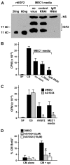
MEC1 and primary epicardial cells secrete IGF2. (A) Western blot. Serum-free media from MEC1 cells was TCA precipitated and evaluated by western blotting. MEC1 cells were treated with no adenovirus, with a control virus expressing an irrelevant sequence (control RNAi), or with virus expressing an RNAi specific for Igf2. Two specific bands for IGF2 (20 kDa and 11 kDa) were detected, plus a 35 kDa nonspecific (NS) band. Recombinant human IGF2 (rhIGF2) was used as a reference for size and quantity. Sizes at left are the migration of standards. (B) Inhibition of Igf2 expression reduces mitogenic activity. NIH3T3 cells were grown in serum-free media (SF), treated with 10% calf serum (CS) as a positive control, or treated with serum-free media isolated from MEC1 cells grown in the absence or presence of the indicated viruses. Proliferation was measured by [3H]thymidine incorporation. (C) Inhibition of IGF signaling reduces mitogenic activity. NIH3T3 cells were grown in the presence of solvent (DMSO) or the IGF receptor inhibitor AG1024 (0.2 μM) in serum-free media (SF), in the presence of 10% calf serum (CS) as a positive control, in the presence of 100 ng/ml recombinant human IGF2, or in serum free media from MEC1 cells. (D) Epicardium-cardiomyocyte co-culture. Embryonic ventricular cardiomyocytes were cultured alone (CM alone) or co-cultured with primary embryonic epicardial cells (CM + epi), and proliferation of Nkx2.5+ cardiomyocytes measured by BrdU incorporation in the absence or presence of AG1024. For all graphs, asterisks indicate statistically significant differences (P<0.05) relative to control treatments and error bars indicate s.d. CPM, counts per minute.
Fig. 3.
Fig. 3.
Expression of IGF components in the developing heart detected by fluorescence in situ hybridization. Fluorescent riboprobes are shown in green, DAPI counterstaining is in blue. Nonspecific red blood cell epifluorescence is seen with varying intensity in all panels. bw, body wall; cz, compact zone; endo, endocardium; epi, epicardium.
Fig. 4.
Fig. 4.
Proliferative consequences of Igf2 and IGF receptor deficiency. (A) Proliferation in the right ventricular wall in control, Igf2-null, and conditional IGF receptor-null embryos at E11.5, visualized by merged three-color fluorescence. Nkx2.5-positive cardiomyocytes appear green, BrdU-positive (proliferating) cardiomyocytes appear orange-yellow, proliferating epicardial or endocardial cells are red and nuclei are blue (DAPI). Individual single color panels are shown in Fig. S5 in the supplementary material. (B,C) Quantification of compact zone cardiomyocyte proliferation in control versus mutant embryos. n=4-5 embryos for each genotype at E11.5 and E12.5, and n=3 at E14.5. Asterisks indicate statistically significant differences (P<0.05) relative to control hearts. Error bars indicate s.d. LV, left ventricle; RV, right ventricle.
Fig. 5.
Fig. 5.
Morphology of embryonic hearts at E14.5. Hematoxylin and Eosin stained sections of hearts of the indicated genotypes; the boxed regions in the low magnification views above are shown at higher magnification below. Corresponding photographs are at the same magnification. The compact zone (cz) is indicated by brackets.
Fig. 6.
Fig. 6.
Inhibition of phosphorylation of ERK in short-term in vitro cultured embryos. E11.5 wild-type embryos were cultured in the presence of solvent (DMSO), the IGFR inhibitor AG1024 (at three different concentrations), the FGFR inhibitor PD173074 (500 nM), or a combination of AG1024 (10 μM) plus PD173074 (500 nM). Protein extracts from the heart (left) or forelimb (right) were analyzed by western blotting for phosphorylated ERK (pErk; above) or total ERK (tErk; below). At the bottom, results from several experiments were combined graphically. The ratio is expressed in arbitrary units (AU) and does not reflect the absolute percentage of pERK; the signal for DMSO-treated (−, negative control) samples was defined as 100 for comparison between experiments. Asterisks indicate statistically significant differences (P<0.05) for heart samples relative to DMSO and to PD173074 treated; for limb samples relative to DMSO and to AG1024 treated. Error bars indicate s.d.

References

    1. Armstrong M. T., Lee D. Y., Armstrong P. B. (2000). Regulation of proliferation of the fetal myocardium. Dev. Dyn. 219, 226-236 - PubMed
    1. Austin A. F., Compton L. A., Love J. D., Brown C. B., Barnett J. V. (2008). Primary and immortalized mouse epicardial cells undergo differentiation in response to TGFbeta. Dev. Dyn. 237, 366-376 - PubMed
    1. Baker J., Liu J. P., Robertson E. J., Efstratiadis A. (1993). Role of insulin-like growth factors in embryonic and postnatal growth. Cell 75, 73-82 - PubMed
    1. Belke D. D., Betuing S., Tuttle M. J., Graveleau C., Young M. E., Pham M., Zhang D., Cooksey R. C., McClain D. A., Litwin S. E., et al. (2002). Insulin signaling coordinately regulates cardiac size, metabolism, and contractile protein isoform expression. J. Clin. Invest. 109, 629-639 - PMC - PubMed
    1. Bondy C. A., Werner H., Roberts C. T., LeRoith D. (1990). Cellular pattern of insulin-like growth factor-I (IGF-I) and type I IGF receptor gene expression in early organogenesis: comparison with IGF-II gene expression. Mol. Endocrinol. 4, 1386-1398 - PubMed

Publication types

MeSH terms

Substances