. 2011 Mar 23;31(12):4434-43.
doi: 10.1523/JNEUROSCI.4874-10.2011.

Recovery from mu-opioid receptor desensitization after chronic treatment with morphine and methadone

Affiliations

Recovery from mu-opioid receptor desensitization after chronic treatment with morphine and methadone

Nidia Quillinan et al. J Neurosci. .

Abstract

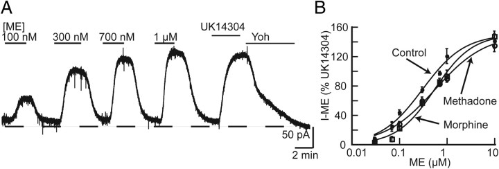
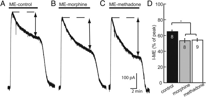
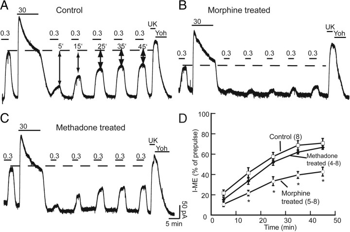
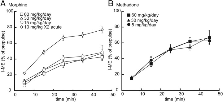
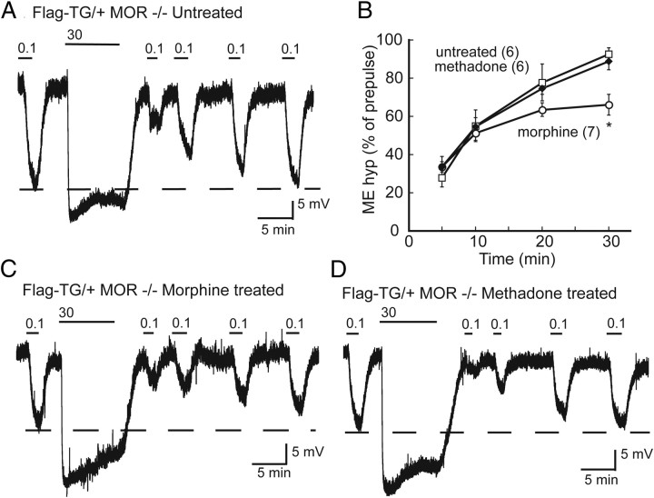
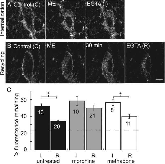
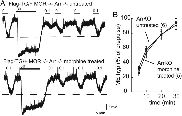
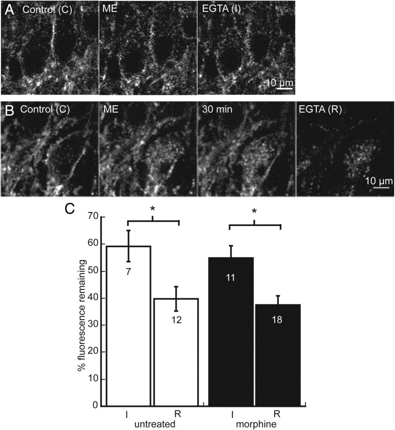
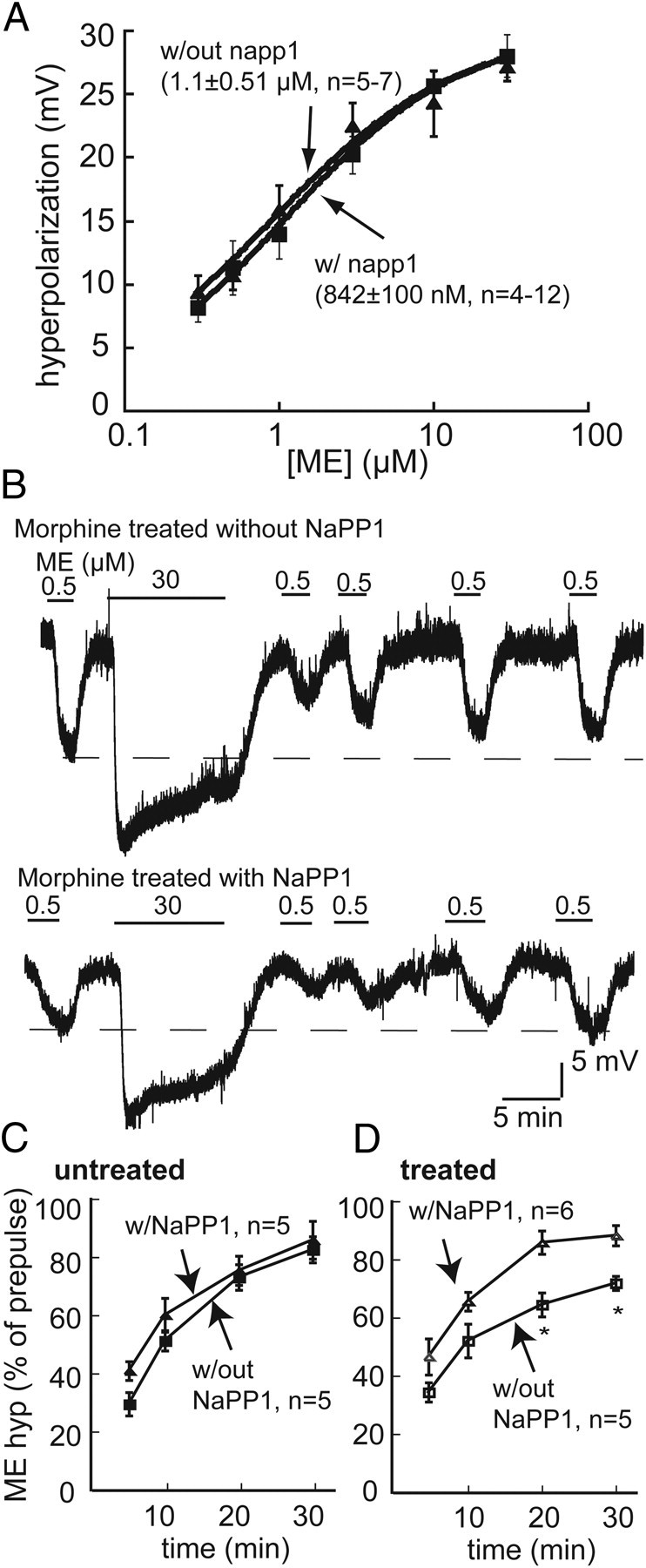
Chronic treatment with morphine results in a decrease in μ-opioid receptor sensitivity, an increase in acute desensitization, and a reduction in the recovery from acute desensitization in locus ceruleus neurons. With acute administration, morphine is unlike many other opioid agonists in that it does not mediate robust acute desensitization or induce receptor trafficking. This study compares μ-opioid receptor desensitization and trafficking in brain slices taken from rats treated for 6-7 d with a range of doses of morphine (60, 30, and 15 mg · kg(-1) · d(-1)) and methadone (60, 30, and 5 mg · kg(-1) · d(-1)) applied by subcutaneous implantation of osmotic minipumps. Mice were treated with 45 mg · kg(-1) · d(-1). In morphine-treated animals, recovery from acute [Met](5)enkephalin-induced desensitization and receptor recycling was diminished. In contrast, recovery and recycling were unchanged in slices from methadone-treated animals. Remarkably the reduced recovery from desensitization and receptor recycling found in slices from morphine-treated animals were not observed in animals lacking β-arrestin-2. Furthermore, pharmacological inhibition of G-protein receptor kinase 2 (GRK2), although not affecting the ability of [Met](5)enkephalin to induce desensitization, acutely reversed the delay in recovery from desensitization produced by chronic morphine treatment. These results characterize a previously unidentified function of the GRK/arrestin system in mediating opioid regulation in response to chronic morphine administration. They also suggest that the GRK/arrestin system, rather than serving as a primary mediator of acute desensitization, controls recovery from desensitization by regulating receptor reinsertion to the plasma membrane after chronic treatment with morphine. The sustained GRK/arrestin-dependent desensitization is another way in which morphine and methadone are distinguished.


Figures

Figure 1.
Figure 1.
Chronic morphine and methadone treatment resulted in reduced potency of ME in rat LC neurons. A, Representative trace showing outward currents produced by perfusion of varying concentrations of ME. At the end of the experiment, the outward current produced by UK14304 (3 μm) was measured and reversed by yohimbine (3 μm; Yoh). B, Concentration–response curves for ME in slices from untreated, chronic morphine (60 mg · kg−1 · d−1) and chronic methadone (60 mg · kg−1 · d−1) treated rats. Outward currents induced by ME (30 nm to 10 μm) were normalized to the peak current induced by UK14304. All points are mean ± SE. The EC50 values from control (281 ± 47 nm), chronic morphine (578 ± 85 nm), and chronic methadone (500 ± 78 nm).
Figure 2.
Figure 2.
Enhanced desensitization observed after chronic treatment of rats with morphine or methadone. A–C, Representative traces showing the decline in the outward current during a 10 min application of a saturating concentration of ME (30 μm). The peak current (dashed line) declined during the 10 min application to a desensitized amplitude (double arrows). D, Bars represent the mean percentage of peak current ± SE in control (black), morphine treated (gray), and methadone treated (white). n is indicated in bars. One-way ANOVA followed by Dunnett's post hoc test showed a significant difference between morphine treated and methadone treated vs control (*p < 0.01).
Figure 3.
Figure 3.
Recovery from desensitization is impaired in slices from mice treated with chronic morphine but not methadone. A, Representative traces of outward currents in slices from untreated mice. ME (300 nm) was perfused before desensitizing with ME (30 μm, 10 min). ME (300 nm) was tested again 5 min after washout of the desensitizing concentration, and the amplitude of the current was compared with the prepulse amplitude (dashed line). ME (300 nm) was tested again at 15, 25, 35, and 45 min to measure recovery from desensitization (percentage of prepulse amplitude). At the end of the experiment, the current induced by UK14304 (3 μm; UK) was determined and reversed with yohimbine (3 μm; Yoh). B, C, Representative traces from morphine-treated and methadone-treated rats. D, Summary of recovery from desensitization (percentage of prepulse amplitude). □, Control; ○, morphine treated; ♦, methadone treated. Error bars represent SEM (n). Two-way ANOVA with Bonferroni's post hoc comparison (*p < 0.001).
Figure 4.
Figure 4.
The recovery from desensitization after morphine treatment of rats was independent of the dose but required more than 1 d of treatment. A, Plot of the recovery from desensitization, as a function of time, in slices taken from rats treated with morphine. When rats were treated for 6–7 d with different doses of morphine, the rate and extent of recovery from desensitization was reduced compared with untreated rats. In slices taken from rats given two injections of morphine on a single day, the recovery from desensitization was not different from untreated animals. B, In slices taken from rats treated with different doses of methadone, the rated and extent of recovery from desensitization was not different from untreated. Error bars represent SEM. Two-way ANOVA with Bonferroni's post hoc comparison (*p < 0.05).
Figure 5.
Figure 5.
Recovery from desensitization impaired after chronic morphine treatment in Flag-TG/+ mice. A, Representative trace of membrane potential in slices from an untreated Flag-TG/+ mouse. ME (100 nm) was perfused before and 5, 10, 20, and 30 min after desensitizing with a saturating concentration of ME (30 μm). After 30 min, recovery from desensitization was nearly complete (dashed line). Representative trace of desensitization and recovery in slices from morphine-treated (B) and methadone-treated (C) mice. D, Summary of recovery from desensitization (percentage of prepulse amplitude). □, Control; ○, morphine treated; ◆, methadone treated. Error bars represent SEM (n). Two-way ANOVA with Bonferroni's post hoc comparison (*p < 0.05).
Figure 6.
Figure 6.
Internalization and recycling of FlagMOR in live brain slices. A, Representative images of ME-induced internalization. Control (C) is initial staining with an Alexa Fluor-594-conjugated anti-Flag M1 antibody before agonist treatment. Internalization was induced with a 10 min perfusion of ME (30 μm). Immediately after washout of ME, calcium-free EGTA solution was perfused to remove M1–A594 from receptors on plasma membrane (I). B, Control (C) image of initial staining. Internalization was induced by ME, and, after a 30 min wash in normal ACSF, some receptors recycled to plasma membrane. Ca-free EGTA solution was then applied to strip antibody from the plasma membrane (R). C, Summarized data of total fluorescence remaining after perfusion of Ca-free EGTA solution immediately after internalization was induced (I/C) or after 30 min (R/C) in slices from untreated (black), morphine-treated (gray,) and methadone-treated (white) mice. Dashed line indicates percentage fluorescence intensity remaining in drug-free experiment. Error bars represent SEM. I/C and R/C were compared within treatment groups using Student's t test (*p < 0.05).
Figure 7.
Figure 7.
Reduced recovery from desensitization in chronic morphine-treated animals requires βArr2; experiments were performed in FlagMOR–βArr2−/− mice. A, Representative traces showing desensitization and recovery in an untreated (top) and morphine-treated (bottom) mouse. Dashed lines represent prepulse amplitude of the hyperpolarization produced by ME (100 nm). B, Summary of recovery (percentage of prepulse amplitude) for untreated (♦) and morphine-treated (•) mice. Two-way ANOVA with Bonferroni's post hoc comparison (*p < 0.05). KO, Knock-out.
Figure 8.
Figure 8.
Internalization and recycling are not altered by chronic morphine treatment in FlagMORArr2−/− mice. A, Internalization induced by ME. Control (C) is initial staining with an Alexa Fluor-594-conjugated anti-Flag M1 antibody before agonist treatment. Internalization was induced with a 10 min perfusion of ME (30 μm) and was followed immediately by Ca-free EGTA to remove M1–A594 from receptors on plasma membrane (I). B, Recycling of receptors after ME-induced internalization. Control (C) is initial staining, and internalization was induced with ME (30 μm). After a 30 min wash in normal ACSF, some receptors recycled to plasma membrane and Ca-free EGTA was applied to remove M1–A594 from receptors on plasma membrane. C, Summarized data of total fluorescence remaining after Ca-free EGTA solution was applied immediately after ME-induced internalization (I/C) and after 30 min wash (R/C) in untreated (white) and morphine-treated (black) Flag-TG/+Arr−/− slices. Error bars represent SEM, and Student's t test was used to compare I/C with R/C within groups (*p < 0.05).
Figure 9.
Figure 9.
Inhibition of GRK2 with 1NaPP1 in the GRK2as5 mice reverses the effects of chronic morphine treatment on recovery from desensitization. A, Concentration–response curve for ME constructed from average membrane hyperpolarizations in slices from GRK2as5 mouse in the absence (■) or presence (▴) of 1NaPP1 (10 μm). B, Representative recordings in slices from morphine-treated GRK2aa5 mice without 1NaPP1 (top) and slices incubated with 1NaPP1 (bottom; 10 μm, 30 min) before desensitizing with ME. C, Summary of recovery (percentage of prepulse amplitude) in slices from untreated GRKas5 without (■) or with (▴) 1NaPP1. D, Summary of recovery in slices from morphine-treated GRKas5 without (□) and with (▵) 1NaPP1. Error bars represent SEM. Two-way ANOVA with Bonferroni's post hoc comparison (*p < 0.05).

Similar articles

Cited by

References

    1. Alvarez VA, Arttamangkul S, Dang V, Salem A, Whistler JL, Von Zastrow M, Grandy DK, Williams JT. mu-Opioid receptors: ligand-dependent activation of potassium conductance, desensitization, and internalization. J Neurosci. 2002;22:5769–5776. - PMC - PubMed
    1. Arttamangkul S, Quillinan N, Low MJ, von Zastrow M, Pintar J, Williams JT. Differential activation and trafficking of micro-opioid receptors in brain slices. Mol Pharmacol. 2008;74:972–979. - PMC - PubMed
    1. Bagley EE, Chieng BC, Christie MJ, Connor M. Opioid tolerance in periaqueductal gray neurons isolated from mice chronically treated with morphine. Br J Pharmacol. 2005;146:68–76. - PMC - PubMed
    1. Bailey CP, Couch D, Johnson E, Griffiths K, Kelly E, Henderson G. μ-opioid receptor desensitization in mature rat neurons: lack of interaction between DAMGO and morphine. J Neurosci. 2003;23:10515–10520. - PMC - PubMed
    1. Bailey CP, Llorente J, Gabra BH, Smith FL, Dewey WL, Kelly E, Henderson G. Role of protein kinase C and μ-opioid receptor (MOPr) desensitization in tolerance to morphine in rat locus coeruleus neurons. Eur J Neurosci. 2009a;29:307–318. - PMC - PubMed

Publication types

MeSH terms