. 2011 Mar 23;31(12):4569-82.
doi: 10.1523/JNEUROSCI.4638-10.2011.

In vivo imaging of dorsal root regeneration: rapid immobilization and presynaptic differentiation at the CNS/PNS border

Affiliations

In vivo imaging of dorsal root regeneration: rapid immobilization and presynaptic differentiation at the CNS/PNS border

Alessandro Di Maio et al. J Neurosci. .

Abstract

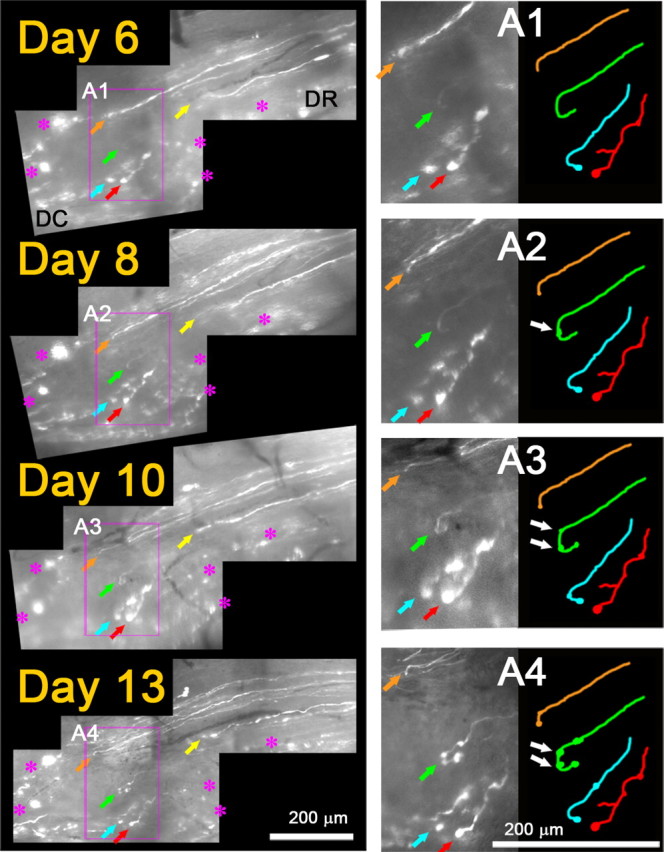
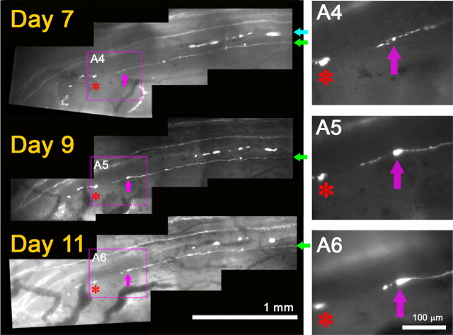
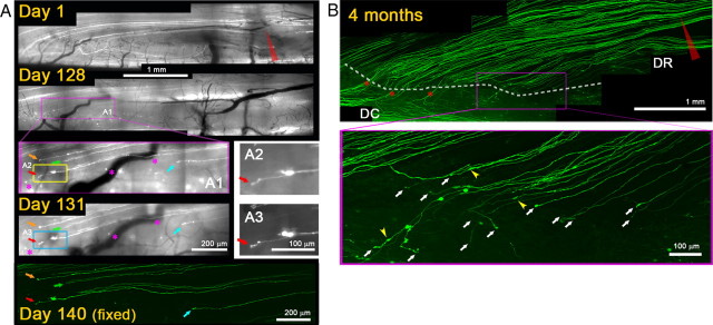
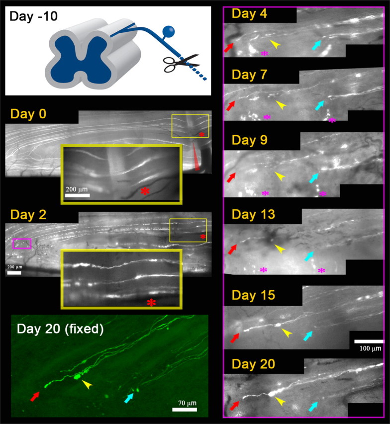
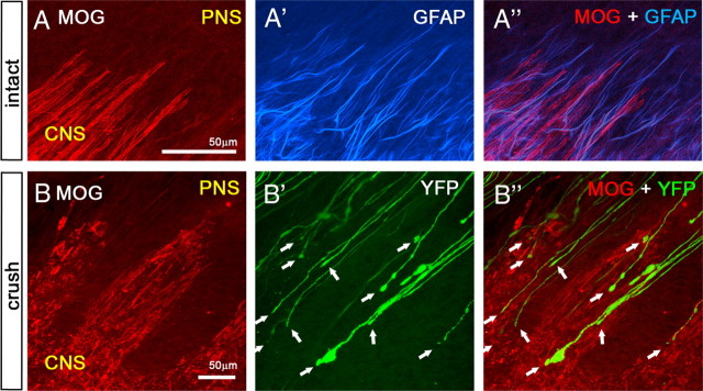
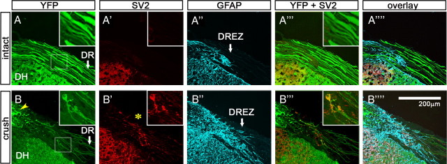
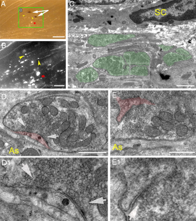
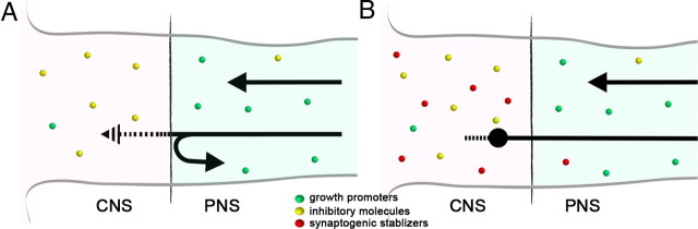
Dorsal root (DR) axons regenerate in the PNS but turn around or stop at the dorsal root entry zone (DREZ), the entrance into the CNS. Earlier studies that relied on conventional tracing techniques or postmortem analyses attributed the regeneration failure to growth inhibitors and lack of intrinsic growth potential. Here, we report the first in vivo imaging study of DR regeneration. Fluorescently labeled, large-diameter DR axons in thy1-YFPH mice elongated through a DR crush site, but not a transection site, and grew along the root at >1.5 mm/d with little variability. Surprisingly, they rarely turned around at the DREZ upon encountering astrocytes, but penetrated deeper into the CNS territory, where they rapidly stalled and then remained completely immobile or stable, even after conditioning lesions that enhanced growth along the root. Stalled axon tips and adjacent shafts were intensely immunolabeled with synapse markers. Ultrastructural analysis targeted to the DREZ enriched with recently arrived axons additionally revealed abundant axonal profiles exhibiting presynaptic features such as synaptic vesicles aggregated at active zones, but not postsynaptic features. These data suggest that axons are neither repelled nor continuously inhibited at the DREZ by growth-inhibitory molecules but are rapidly stabilized as they invade the CNS territory of the DREZ, forming presynaptic terminal endings on non-neuronal cells. Our work introduces a new experimental paradigm to the investigation of DR regeneration and may help to induce significant regeneration after spinal root injuries.


Figures

Figure 1.
Figure 1.
YFP-labeled DRG neurons are large and neurofilament positive. A, Size distribution of YFP+ L5 DRG neurons (green) in Thy1-YFPH mice, superimposed on the entire population, labeled with a fluorescent Nissl stain (red, inset). YFP+ neurons are large. B, Superficial layers of the dorsal horn of a Thy1-YFPH mouse, exhibiting little YFP fluorescence in lamina II, where CGRP+ and IB4+ innervation is abundant. Yellow dotted lines denote dorsal roots. C, Four-color immunolabeling of a DRG cross section illustrating expression of neurofilament (magenta) but not CGRP (blue) or IB4 (red) in YFP+ DRG neurons (green) in Thy1-YFPH mice. Scale bars: A, 200 μm; B, 200 μm; C, 50 μm.
Figure 2.
Figure 2.
Axons fail to elongate through a transection injury. A, Schematic illustration of a right-sided laminectomy (T12–L5) to partially expose the L5 root. B, Low-magnification fluorescence view of the exposed spinal cord of a living Thy1-YFPH mouse. The laminectomy extends laterally to partially expose L3, L4, and L5 DRGs and medially to expose the midline dorsal vein. C, High-magnification fluorescence view of the area where L5 DR axons enter the spinal cord. Superficially positioned YFP+ axons run parallel to the midline dorsal vein, curve perpendicularly to enter the DREZ, and then bifurcate within the spinal cord. Highlighted box approximates DREZ. D, Repeated imaging of L5 DR axons for 7 d after root transection. Axons in L5 root were cut near the L3 DRG (red scissors), ∼3 mm from the DREZ, and imaged on day 0, day 3 (not shown), and day 7. The area of the root transection (red boxes) is magnified in right panels (D1–D3). D1, Before the transection. A few superficial YFP+ axons are visible. D2, After the transection. The medial portion of the L5 root was completely cut with spring scissors, and the proximal and distal ends were then closely reapposed. D3, Seven days after the transection. The transection site is filled with ∼500 μm collagenous scar tissue. No axons cross it. D4, A high-magnification confocal view of the area prepared after sacrificing the mouse on day 7. YFP+ axons located deeper in the root than those imaged in vivo also failed to regenerate across the transection site (asterisks). Instead, they turned around (arrows) and extended back along the proximal axons. Note that all the fluorescence in the stump beyond the cut (i.e., left of the asterisk) is due to fragments of degenerating axons that retained YFP.
Figure 3.
Figure 3.
Axons elongate through a crush injury with little variability. A, Repeated imaging of L5 DR axons for 7 d after root crush. The medial portion of the L5 root was crushed with a fine forceps (red arrowhead) and imaged on days 0, 3, 5, and 7 after the crush. The area of the crush is magnified in the right panels (A1–A4). A1, Immediately after crush. Four injured YFP+ axons are shown (colored arrows). A2, Three days after the crush. All four axons could be reidentified. Each axon extends a single neurite that crosses the site of crush. A3, A4, Five and seven days after crush. Neurites remain stable and there is no additional growth from these or other proximal axons. Axon swellings are occasionally observed (e.g., A4; axon marked by blue arrows). A5, High-magnification confocal view of the area prepared after sacrificing the mouse on day 7. YFP+ axons located deeper in the root also regenerate across the crush site. B, Confocal analysis of the regeneration after a crush injury in another Thy1-YFPH mouse that did not receive in vivo imaging. The L5 root was crushed at the usual location (red arrowhead); 4 d later, the spinal cord was prepared in whole mounts and analyzed by high-resolution confocal microscopy. The area near the crush injury (blue box) and the DREZ (yellow box) on day 4 are magnified in B1 and B2, respectively. B1, Consistent with in vivo imaging observations, almost all YFP+ axons extended a single neurite that crossed the crush site and grew further without stopping. Blue arrows point to degenerating axon fragments. B2, Almost all axons reached the DREZ and terminated at a location similar to that of single axons (arrows). Few axons turned around (e.g., arrowhead), further evidence of the consistency of the axon response to crush injury. DC, Dorsal column; DR, dorsal root. Red stars indicate uninjured axons. Because their proximal portion was located laterally, they escaped crush of the medial portion of the L5 root.
Figure 4.
Figure 4.
Axons elongate >1.5 mm/d along preexisting endoneurial tubes. A, Repeated imaging of an identified axon for 4 d after root crush. On day 0, a Thy1-YFPH mouse with only a few superficial YFP+ axons underwent crush of the most medial portion of the L5 root (red arrowheads) to minimize the number of damaged axons. A1, Magnified view of the crush site immediately after injury. Two superficial axons are shown; the axon marked by a blue arrow (blue axon) was stretched but survived the injury, whereas the axon marked by a green arrow (green axon) was damaged. On day 2, degeneration of both proximal and distal tips confirmed apparent damage of the green axon by the crush. A2, Magnified view of the crush site. Yellow arrow denotes dying-back degeneration of the green axon. Note that no neurite has yet been formed by the green axon. On day 4, the green axon has extended ∼3 mm, and its tip has reached the DREZ. A3, Magnified view of the green axon near the DREZ, illustrating its growth along the fluorescent fragments of degenerating distal axons (yellow arrowheads: i.e., endoneurial tube trajectory). B, Examples of newly formed neurites on proximal stump axons imaged 2 d after injury. Day 0, Magnified view of the crush site immediately after injury showing two crushed axons (pink arrows). Day 2, Both axons extended short neurites (green arrows) that have not yet reached the distal stump axons (e.g., yellow arrowhead). These neurites have slender growing tips (green arrow, inset). Day 3, Both neurites extended through the degenerating distal axons. Asterisks in A1 and A2 indicate a node of Ranvier on the blue axon that served as a landmark. Blood vessels served as a landmark in B. Red arrowheads indicate the site of crush.
Figure 5.
Figure 5.
Regenerating axons are rapidly immobilized at the DREZ. Repeated imaging of axon tips at the DREZ 6–13 d after root crush. On day 0 (data not shown), the L5 root was crushed at the usual site, ∼3 mm away from the DREZ. On day 6, tips of five regenerating axons are observed at the DREZ (colored arrows). DC, Dorsal column; DR, dorsal root. Magenta asterisks denote fluorescent debris of degenerating old axons that served as landmarks. Note that the location of an axon tip relative to other axon tips and landmarks remained unchanged in subsequent imaging sessions on days 8, 10, and 13, indicating that the axons were immobilized after entering the DREZ. A1–A4, Magnified view of the area outlined by magenta boxes showing tips of four axons. They did not grow, retract, or turn around and remained in the same location in subsequent imaging sessions. Their appearance also was unchanged except for swellings that developed on the tips or shafts of some axons (e.g., white arrows in A2–A4).
Figure 6.
Figure 6.
Immobilized axon endings at the DREZ extend and retract a sprout. Shown is continued imaging of the mouse shown in Figure 4, 7–11 d after L5 root crush. On day 7, the tip of the green axon (magenta arrows) is at the same location as on day 4 (cf. Fig. 4). The blue axon that was stretched on day 0 has degenerated. Red asterisks indicate axon debris, a landmark, that remained stable during the imaging sessions. Note that the relative distance between the tip of the green axon and the debris remained the same on subsequent imaging sessions on days 9 and 11. A4, Magnified view of the axon tip on day 7. A fine neurite extends from the slightly swollen tip (magenta arrow). A5, Two days later on day 9, the neurite has elongated ∼50 μm; the axon tip remains at the same location and is more swollen. A6, Two days later on day 11, the neurite appears to have absorbed into the tip, which remains immobilized at the same location. Thus, the apparent mobility of this axon at the DREZ was due to fruitless sprouting of a stabilized axon tip.
Figure 7.
Figure 7.
Axons are chronically immobilized at the DREZ. A, Repeated imaging of axons crushed 4 months previously. L5 root of a Thy1-YFPH mouse was crushed (red arrow) and imaged on days 1, 128, 131, and 140 following injury. On day 1, the root crush is indicated by degeneration of proximal and distal axon stumps at the injury site. On day 128, several axons are located at the DREZ outlined by a magenta box. A1, Magnified view of the DREZ area showing at least five axons and their tips (colored arrows). A2, Magnified view of the yellow boxed area in A1, showing an axon and its tip marked by red arrows. The location and appearance of these axons and their tips remained unchanged on subsequent imaging sessions on days 131 and 140. A3, Magnified view of the axon in A2 on day 131, illustrating its chronic stability. The day 140 image is a confocal view of the area prepared in a fixed whole mount. B, Confocal analysis of a different mouse that was not imaged in vivo. Top, L5 root was crushed at the usual location (red arrowhead) and, 4 months later, the spinal cord was analyzed in a whole-mount preparation. White dotted line, Location of axon tips at the DREZ. Asterisks, Intact axons that were uninjured because of their location lateral to the crush site. Bottom panel, Magnified view of the magenta-boxed area. Almost no axons turned around, and their tips (white arrows) were found at a location similar to those in mice imaged in vivo on day 140 (cf. Fig. 7A) and on day 4 (cf. Fig. 3B2) after root crush, further evidence of the chronic stability of axons at the DREZ. Yellow arrowheads, Swellings formed on axon shafts.
Figure 8.
Figure 8.
Axons are rapidly immobilized at the DREZ even after conditioning lesion. Repeated imaging of conditioning lesioned DR axons over 20 d after L5 root crush. Day −10, A schematic drawing illustrating a conditioning lesion of the ipsilateral sciatic nerve 10 d before DR crush. Day 0, Immediately after the root crush, 10 d after conditioning lesion. L5 root was crushed at the usual location (red arrowhead). Three crushed axons are shown in the large yellow inset box that magnifies the superficial site of crush (small yellow box). Day 2, All three axons have already extended neurites across the crush site, illustrating enhanced growth within the root due to a conditioning lesion. Magenta box, An area of the DREZ where several axons were monitored in subsequent imaging sessions, presented in magnified views in the right panels. Day 4, No axons regenerated through the DREZ (data not shown). The tips of these axons remain in the same location and have a similar appearance in subsequent imaging sessions on days 7, 9, 13, 15, and 20. Positions of an axon tip relative to other axon tips and landmarks were used to determine axon motility between imaging sessions. Yellow arrowhead denotes the tip of an axon with a particularly large increase in size over time. These axons were found again after the mouse was killed, and high-resolution confocal microscopy confirmed the location and appearance of the axon tips [day 20 (fixed)].
Figure 9.
Figure 9.
Axons terminate in CNS territory containing oligodendrocytes. A–A″, Low-magnification confocal view of the glial interface at the DREZ in an intact Thy1-YFPH animal. Oligodendrocytes (A, red) and astrocytes (A′, blue) were labeled with MOG and GFAP antibodies, respectively, in a whole-mount preparation. Note that astrocytes extend further into the periphery than oligodendrocytes even in intact, noninjured DREZ (A″). Axons (green) were omitted. B–B″, Confocal view of the glial interface at the injured DREZ. The L5 root was crushed 2 weeks previously. Oligodendrocytes are degenerating (B, red), whereas astrocytes invade further into the PNS (data not shown) (cf. Fig. 10A″,B″). Axons (B′, YFP) do not stop when they encounter astrocytic processes in the interface (data not shown) but terminate deeper in CNS territory containing oligodendrocytes (B″, white arrows).
Figure 10.
Figure 10.
Intense immunoreactivity of synapse markers associated with axon tips and shafts at the DREZ. Cross sections of the cervical roots of intact (A–A″″) and crushed (B–B″″) Thy1-YFP16 mice were immunolabeled with antibodies against SV2 (red) and GFAP (blue). The cervical roots (C3–C5) of the injured mouse were crushed peripherally 20 d previously. A, In the intact Thy1-YFP16 mouse, all of the large-diameter DR axons were labeled. DR, Dorsal root; DH, dorsal horn. A′, SV2 immunoreactivity is not observed along the DR or at the DREZ. A″, GFAP-labeled astrocytes denote the glial interface at the DREZ (arrow). No SV2 (A′) or synaptotagmin (data not shown) immunoreactivity is present in the CNS territory of the DREZ (A″″–A″″). Insets magnify an area of the DREZ. B, In the injured mouse, numerous regenerating axons stop at the DREZ. The inset is a magnified view of an area of the DREZ showing two axons and their tips. Yellow arrowhead indicates debris of degenerating YFP+ axons. B′, Intense SV2 immunoreactivity is present at the DREZ where axons terminate. The inset illustrates intense SV2 immunoreactivity associated with the two axon tips and shafts. B″, Astrocytes invade further into the periphery in the injured mouse. Note that SV2 immunoreactivity associated with the axon tips and shafts is present in the CNS territory as marked by GFAP-labeled astrocytes (B″″, B″″).
Figure 11.
Figure 11.
Ultrastructural analysis of DR axons stopped at the DREZ, revealing presynaptic differentiation. The L5 root of a Thy1-YFPH mouse was crushed 13 d previously. A, Low-magnification, transmitted-light view, superimposed on fluorescence image, of the DREZ in a vibratome slice. Photoconverted DiI crystals (white arrows) were placed in an area where axons were stopped, and the area between the two crystals was examined in the transmission electron microscope. Yellow arrowheads point to two axon tips. The asterisk indicates axon debris and marks the same spot in B. B, Magnified fluorescent view of the boxed area in A. Axons stopped at the usual location in the DREZ, showing axonal debris (asterisk) and stalled axon tips (yellow arrowheads). C, An electron micrograph of the targeted area of the DREZ showing axonal profiles (pseudocolored green) embedded within non-neuronal cellular processes. D, Magnified view of a boxed area in C, revealing a nerve-terminal-like profile in contact with a non-neuronal cellular process (pseudocolored red). Note that the presynaptic profile is filled with differentially distributed mitochondria and abundant ∼40 nm vesicles but lacks vacuoles and disorganized microtubules. D1, An enlarged area of synaptic contact in D. Vesicles are highly clustered and docked at an electron-dense membrane that resembles an active zone (white arrows). No postsynaptic densities are present on the non-neuronal cell process. E, Magnified view of another area in C, revealing a nerve-terminal-like profile of an adjacent axon. E1, An enlarged area of synaptic contact in E. Note that this axon also shows presynaptic differentiation in contact with a fine non-neuronal cellular process (pseudocolored red) that does not exhibit postsynaptic densities. SC, Schwann cell; As, astrocyte. Scale bars: A, 250 μm; B, 100 μm; C, 1 μm; D, 250 nm; E, 200 nm.
Figure 12.
Figure 12.
Schematic illustration of the current and proposed models of regeneration failure at the DREZ. A, The model that illustrates the prevailing view in the field. Black arrows represent regenerating DR axons. Sensory axons lack intrinsic growth potential but are capable of regenerating in the PNS due to abundant growth-promoting molecules (green circles). Their growth is prevented at the DREZ by growth inhibitors abundant in the CNS (yellow circles). According to this model, many axons must be repelled at the CNS/PNS interface (i.e., an arrow turned around to the PNS). Axons that invade CNS territory must be prompted to retract (i.e., patterned arrow) into the PNS by growth inhibitors that transiently collapse growth cones and then either attempt to reenter the CNS or grow further into the dorsal root. This model fails to explain rapid, long-lasting immobilization of most of the axons in the CNS territory of the DREZ. B, Proposed model. In addition to growth inhibitors (yellow circles), the CNS territory of the DREZ is enriched with synaptogenic molecules (red circles). Growth inhibitors alone are not powerful enough to repel most axons at the interface. Accordingly, most axons invade the CNS territory of the DREZ. They are, however, immobilized rapidly by synaptogenic activity that induces presynaptic differentiation (i.e., swollen axon ending). Both growth inhibitors and synaptogenic stabilizers prevent further growth or fruitless sprouting of the immobilized axon tips (i.e., patterned line protruded from the swollen axon ending).

References

    1. Adcock KH, Brown DJ, Shearer MC, Shewan D, Schachner M, Smith GM, Geller HM, Fawcett JW. Axon behaviour at Schwann cell–astrocyte boundaries: manipulation of axon signalling pathways and the neural adhesion molecule L1 can enable axons to cross. Eur J Neurosci. 2004;20:1425–1435. - PubMed
    1. Allen NJ, Barres BA. Neuroscience: glia—more than just brain glue. Nature. 2009;457:675–677. - PubMed
    1. Andrews MR, Czvitkovich S, Dassie E, Vogelaar CF, Faissner A, Blits B, Gage FH, ffrench-Constant C, Fawcett JW. α9 integrin promotes neurite outgrowth on tenascin-C and enhances sensory axon regeneration. J Neurosci. 2009;29:5546–5557. - PMC - PubMed
    1. Beggah AT, Dours-Zimmermann MT, Barras FM, Brosius A, Zimmermann DR, Zurn AD. Lesion-induced differential expression and cell association of Neurocan, Brevican, Versican V1 and V2 in the mouse dorsal root entry zone. Neuroscience. 2005;133:749–762. - PubMed
    1. Bergles DE, Roberts JD, Somogyi P, Jahr CE. Glutamatergic synapses on oligodendrocyte precursor cells in the hippocampus. Nature. 2000;405:187–191. - PubMed

Publication types

MeSH terms

Substances

LinkOut - more resources