. 2011 May 1;186(9):5244-53.
doi: 10.4049/jimmunol.1004040. Epub 2011 Mar 23.

Analysis of the role of IL-21 in development of murine B cell progenitors in the bone marrow

Affiliations

Analysis of the role of IL-21 in development of murine B cell progenitors in the bone marrow

Nathalie Simard et al. J Immunol. .

Abstract

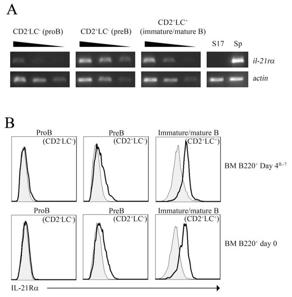
IL-21 plays a key role in the late stage of B cell development, where it has been shown to induce growth and differentiation of mature B cells into Ig-secreting plasma cells. Because IL-21R has also been reported on bone marrow (BM) B cell progenitors, we investigated whether IL-21R influenced earlier stages of B cell development. IL-21R is functional as early as the pro-B cell stage, and the strength of receptor-mediated signaling increases as cells mature. The addition of IL-21 to B cell progenitors in cell culture resulted in the accelerated appearance of mature B cell markers and was associated with the induction of Aid, Blimp1, and germline transcripts. We also found that stimulation of both IL-21R and CD40 was sufficient to induce the maturation of early B cell progenitors into IgM- and IgG-secreting cells. Consistent with a role for IL-21 in promoting B cell differentiation, the number of B220(+)CD43(+)IgM(-) pro-B cells was increased, and the number of mature IgM(hi)IgD(hi) cells was decreased in BM of IL-21R-deficient mice. We also report in this paper that IL-21 is expressed by BM CD4(+) T cells. These results provide evidence that IL-21R is functional in B cell progenitors and indicate that IL-21 regulates B cell development.


Conflict of interest statement

Disclosures

The authors have no financial conflict of interest.

Figures

FIGURE 1
FIGURE 1
IL-21 is produced by CD4+ T cells in BM. A, RT-PCR was performed on cDNA obtained from BM cells of WT, Rag−/− and TCRβ−/− mice. B, Freshly isolated BM or BM B220 cells from WT mice were stained with anti-B220, -CD3, -CD4, -CD8, and -NK 1.1 Abs, and sorted into B cells (B220+), CD4+ T cells (B220CD3+CD4+NK1.1), CD8+ T cells (B220CD3+CD8+NK1.1), and NK cells (B220NK1.1+). RNA was extracted and RT-PCR was performed using primers specific for Il21 and Actin as described in “material and methods”. Spleen was used as a positive control. C, Freshly isolated BM cells from WT mice were stained with anti-CD3, -CD4, -CD44, and –CD69, and sorted into naïve T cells (CD3+CD4+CD44loCD69) and memory T cells (CD3+CD4+CD44hiCD69) populations. Il21 mRNA expression was measured by Real-time PCR and normalized to β-Actin. Spleen was used as a positive control. The figure is a representative experiment of two independent experiments. D, BM CD4+ cells were sorted and cultured with anti-CD3 (10 μg/mL), anti-CD28 (2 μg/mL) with IL-6 (100ng/mL) (solid line) and without IL-6 (100 ng/mL) (dashed line). After 72 hours, IL-21 protein was detected in the supernatant by FACS with beads array. Data presented is a representative experiment that was performed two times with similar results.
FIGURE 2
FIGURE 2
IL-21R expression on BM B cell progenitors. A, Day 4IL-7 B220+ BM cells were stained with anti-CD2, -κ, and -λ Abs, and sorted into proB (CD2LC), preB (CD2+LC), and immature/mature (CD2+LC+) B cell populations. RNA was extracted and semi-quantitative RT-PCR was performed on serial dilutions of cDNA (undiluted, 1/5 dilution, 1/10 dilution) using primers specific for IL-21R and actin as described in the “materials and methods”. The stromal cell line S17 was used as negative control, and spleen (Sp) cells were used as positive control. Semi-quantitative RT-PCR was performed on four independent sample sets with similar results. B, Surface expression of IL-21R on B cell progenitors. Top panel – Day 4IL-7 B220+ BM cells from WT mice and IL-21R−/− mice were stained with anti-B220, -CD2, -kappa LC, -lambda LC, and –IL-21Rα. Bottom panel - Freshly isolated BM cells from WT mice and IL-21R−/− mice were stained with anti-CD2, -κ, -λ, and -IL-21Rα Abs and analyzed by FACS. Open and closed histograms represent binding of anti-mouse IL-21Rα Ab in WT and IL-21R−/− mice, respectively. Data are representative of two independent experiments.
FIGURE 3
FIGURE 3
IL-21R stimulation increases tyrosine phosphorylation of STAT1, STAT3 and STAT5 in proB, preB and immature/mature B cells. Day 4IL-7 B220+ BM cells from WT mice were sorted for proB (CD2LC), preB (CD2+LC) and immature/mature B (CD2+LC+) cells. A, Tyrosine-phosphorylated forms of STAT1 (pY701), STAT3 (pY705), and STAT5 (pY694) were detected by Western blotting. Actin represents loading control. B, The IL-21 non-responsive B62c proB cell line was spiked with different numbers of sorted preB cells, stimulated for 15 min. with IL-21 (50 ng/ml), and analyzed for STAT3 phosphorylation by Western blotting. C, B220+ BM cells were isolated from RAG−/− mice and grown in IL-7. On day 4, cells were harvested and stimulated for 15 min with IL-21 (50 ng/ml). Phosphorylation of STAT3 was detected by Western blotting. D, Day 4IL-7 BM B220+ cells from WT and IL-21R−/− mice were sorted into proB (CD2LC), preB (CD2+LC) and immature/mature B (CD2+LC+) populations. Phosphorylation of STAT3 was detected by Western blotting. E, Day 4IL-7 BM B220+ cells from WT and IL-21R−/− mice were simulated with IL-7 or IL-21. Phospho-STAT3 was detected by Western blotting. Data are representative of two independent experiments.
FIGURE 4
FIGURE 4
IL-21 regulates maturation of different B cell progenitors. Day 4IL-7 B220+ BM cells from WT and IL-21R−/− mice were sorted into proB (CD2LC), preB (CD2+LC) and immature/mature B CD2+LC+) cell populations. Each population was plated with or without IL-21 (30 ng/ml) for 48 hours. Control and IL-21-containing cell cultures initiated with proB (A and B) and preB (C and D) cells from IL-21R−/− and WT mice were harvested on day 2 of culture and analyzed by FACS using CD2-LC and IgM-IgD Ab combination. AC), Percentages of cells in each population are shown on the plots of a representative experiment. BD), Scatter plot of cell population percentages from separate experiments. Lines connect pairs of experiments performed. Statistical analysis of percentage of preB and immature/mature B cells obtained in control and IL-21-treated WT cell cultures was assessed by 2-tailed paired Student t-tests. The analysis shows that there were a significant increase percentage of preB cells in IL-21 containing culture started with proB cells compare to control (p < 0.0001*) (B; lower panel), and a trend toward an increase percentage of immature/mature B cells in IL-21-containing culture initiated with preB cells (percentage of CD2+LC+, p=0.09 (D; lower left panel); percentage of CD2+LChi, p=0.08) (D; lower right panel).
FIGURE 5
FIGURE 5
IL-21R−/− mice have more proB and fewer mature B cells than WT mice in BM. BM cells were isolated from WT (n=20) and IL-21R−/− (n=20) mice and stained for B220, CD43, and IgM; or CD19, CD2, and IgM; or CD19, IgM, and IgD. Percentages (A) and absolute numbers (B) are shown for each cell population. Statistical significance was assessed by 2-tailed Student t test and the level of significance was established at p < 0.05.
FIGURE 6
FIGURE 6
IL-21 regulates gene expression of Blimp1 and Aid in B cell progenitors. B220+ BM cells were grown in IL-7. On day 3, IL-21 (30 ng/mL) was added for 24 hours to half of the culture. Cells were sorted on day 4 into proB (CD2LC), preB (CD2+LC) and immature/mature B (CD2+LC+) cells and total RNA was extracted. Real-time PCR analysis using Blimp1 (A) and Aid (B) specific-primers were performed on cDNA as described in “materials and methods”. Figures show normalized values from a single experiment (left panel) and fold change over untreated from two independent experiments (right panel).
FIGURE 7
FIGURE 7
IL-21R stimulation induces the expression of γ2b germline transcripts. A, Day 4IL-7 BM cells were sorted into proB (B220+CD2LC), preB (B220+CD2+LC), and immature/mature B (B220+CD2+LC+) cells. Each population was plated with IL-21 (30ng/mL), with anti-CD40 (2ug/ml) or anti-CD40 and IL21. After 24 hours, total RNA was extracted and RT-PCR analysis was performed to detect GLTγ2b. β-Actin was used to normalize for cDNA input. Band intensity was measured and normalized to β-Actin using Quantity One software. The figure shows one representative experiment that has been performed at least three times with similar results. In pre-B cells stimulated with IL21, the fold increase in GLT levels ranged from 1.12 to 3.11 fold; in immature/mature B cells, the fold increase ranged from 1.23 to 6.25. Statistical analysis of quantitated and normalized band intensity for pre B cells was performed using the Wilcoxian test demonstrating that there was a significant fold increase with IL21 treatment in preB (CD2+LC) sorted cells (p=0.032, n=6). B, IL-21 in combination with anti-CD40 induces the formation of Ig secreting cells from cultures initiated from B cells progenitors. Left panel, Day 4IL-7 B220+ BM cells were plated with IL-7 in combination with anti-CD40, IL-21 or both. Supernatants were harvested on day 7 and ELISA was performed as described in “materials and methods”. Right panel, Day 4IL-7 B220+ BM cells were sorted into proB cells (CD2LC) and plated with IL-7 and anti-CD40 with or without IL-21. Culture supernatants were collected on day 9 and ELISA was performed as described in “materials and methods”. Results are representative of three independent experiments.

Similar articles

Cited by

References

    1. Lu L, Chaudhury P, Osmond DG. Regulation of cell survival during B lymphopoiesis: apoptosis and Bcl-2/Bax content of precursor B cells in bone marrow of mice with altered expression of IL-7 and recombinase-activating gene-2. J Immunol. 1999;162:1931–1940. - PubMed
    1. Sudo T, Ito M, Ogawa Y, Iizuka M, Kodama H, Kunisada T, Hayashi S, Ogawa M, Sakai K, Nishikawa S. Interleukin 7 production and function in stromal cell-dependent B cell development. J Exp Med. 1989;170:333–338. - PMC - PubMed
    1. Corcoran AE, Smart FM, Cowling RJ, Crompton T, Owen MJ, Venkitaraman AR. The interleukin-7 receptor alpha chain transmits distinct signals for proliferation and differentiation during B lymphopoiesis. Embo J. 1996;15:1924–1932. - PMC - PubMed
    1. Miller JP, Izon D, DeMuth W, Gerstein R, Bhandoola A, Allman D. The earliest step in B lineage differentiation from common lymphoid progenitors is critically dependent upon interleukin 7. J Exp Med. 2002;196:705–711. - PMC - PubMed
    1. Fleming HE, Paige CJ. Pre-B cell receptor signaling mediates selective response to IL-7 at the pro-B to pre-B cell transition via an ERK/MAP kinase-dependent pathway. Immunity. 2001;15:521–531. - PubMed

Publication types

MeSH terms