Randomized Controlled Trial
. 2011 Jun;36(7):1466-77.
doi: 10.1038/npp.2011.32. Epub 2011 Mar 23.

Pregabalin influences insula and amygdala activation during anticipation of emotional images

Affiliations
Randomized Controlled Trial

Pregabalin influences insula and amygdala activation during anticipation of emotional images

Robin L Aupperle et al. Neuropsychopharmacology. 2011 Jun.

Abstract

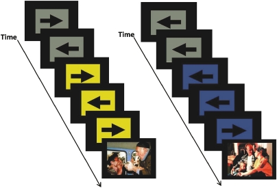
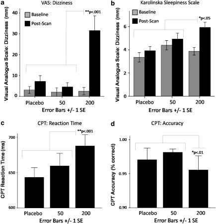
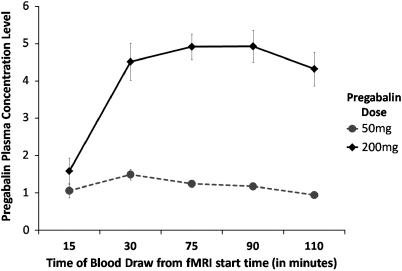
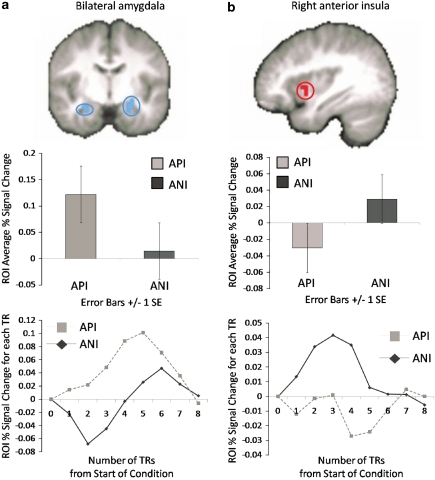
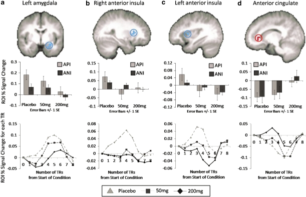
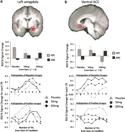
Pregabalin (PGB) has shown potential as an anxiolytic for treatment of generalized and social anxiety disorder. PGB binds to voltage-dependent calcium channels, leading to upregulation of GABA inhibitory activity and reduction in the release of various neurotransmitters. Previous functional magnetic resonance imaging (fMRI) studies indicate that selective serotonin reuptake inhibitors and benzodiazepines attenuate amygdala, insula, and medial prefrontal cortex activation during anticipation and emotional processing in healthy controls. The aim of this study was to examine whether acute PGB administration would attenuate activation in these regions during emotional anticipation. In this double-blind, placebo-controlled, randomized crossover study, 16 healthy controls completed a paradigm involving anticipation of negative and positive affective images during fMRI approximately 1 h after administration of placebo, 50, or 200 mg PGB. Linear mixed model analysis revealed that PGB was associated with (1) decreases in left amygdala and anterior insula activation and (2) increases in anterior cingulate (ACC) activation, during anticipation of positive and negative stimuli. There was also a region of the anterior amygdala in which PGB dose was associated with increased activation during anticipation of negative and decreased activation during anticipation of positive stimuli. Attenuation of amygdala and insula activation during anticipatory or emotional processing may represent a common regional brain mechanism for anxiolytics across drug classes. PGB induced increases in ACC activation could be a unique effect related to top-down modulation of affective processing. These results provide further support for the viability of using pharmaco-fMRI to determine the anxiolytic potential of pharmacologic agents.


Figures

Figure 1
Figure 1
Functional magnetic resonance imaging (fMRI) anticipation task. This task combines a continuous performance task with the interspersed presentation of affective stimuli. Subjects are asked to press a left or right button based on the direction of the arrow. Subjects are instructed before the task that a blue square accompanied by a low tone indicates a positive image is going to appear. In contrast, a switch to a yellow square accompanied by a high tone signals an impending negative image. Images used in this paradigm were taken from the International Affective Picture System (IAPS) (Lang et al, 2008). The color reproduction of this figure is available on the html full text version of the manuscript
Figure 2
Figure 2
Pregabalin (PGB) dose effect on self-report and behavioral measures. PGB exhibited dose effects on self-report measures related to (a) increased dizziness as measured by visual analogue scale (post hoc tests revealed (0=50)<200), (b) increased sleepiness as measured by the Karolinska Sleepiness Scale (KSS; Akerstedt and Gillberg, 1990; post hoc tests revealed (0=50)<200). PGB also exhibited effects on performance during the continuous performance task (CPT; averaged across conditions) conducted during the functional magnetic resonance imaging (fMRI) anticipation paradigm, including (c) increased reaction time (post hoc tests revealed (0=50)>200) and (d) decreased accuracy (post hoc tests revealed (0=50)<200).
Figure 3
Figure 3
Pregabalin plasma concentration levels at various time points during functional magnetic resonance imaging (fMRI) scanning. Plasma was assessed at National Medical Services Lab using high-performance liquid chromatography. Both 50 and 200 mg was associated with detectable plasma concentration levels and greater dose was associated with significantly greater concentration levels.
Figure 4
Figure 4
Task valence effect on functional magnetic resonance imaging (fMRI) blood oxygenation level-dependent (BOLD) activation. Main effects for valence of anticipation, regardless of PGB dose, were observed for (a) bilateral amygdala (shown at y=−8), which exhibited less activation (% signal change) for negative anticipation (ANI) than positive anticipation (API) and (b) right anterior insula (shown at x=36), which exhibited greater activation for ANI than API. Graphs depicted below each image represent (1) average percent signal change (PSC) for each condition and (2) average PSC from the start of the anticipation conditions at each TR time point for each condition.
Figure 5
Figure 5
Pregabalin (PGB) dose effect on functional magnetic resonance imaging (fMRI) blood oxygenation level-dependent (BOLD) activation. PGB attenuated activation within in (a) left amygdala (shown at y=4; post hoc tests revealed (PLB=50 mg)>200 mg) and (b, c) bilateral anterior insula (right insula/claustraum shown at x=28 and left insula shown at x=−34; post hoc tests revealed PLB>(50 mg=200 mg)) and increased activation in (d) anterior cingulate (ACC; shown at x=14; post hoc t-tests revealed (PLB=50 mg)>200 mg), for anticipation of negative (ANI) and positive images (API). Graphs depicted below each image represent (1) average percent signal change (PSC) for each condition and (2) average PSC from the start of the anticipation conditions at each TR time point for each PGB dose (averaged across negative and positive conditions).
Figure 6
Figure 6
Pregabalin (PGB) dose × valence interaction effect on functional magnetic resonance imaging (fMRI) blood oxygenation level-dependent (BOLD) activation. Increasing PGB dose was associated with increased activation to anticipation of negative (ANI) images and decreased activation to anticipation of positive images (API) within the (a) left amygdala (shown at y=2; post hoc tests revealed PLB>50 mg>200 mg) and (b) ventral ACC (shown at x=3; post hoc tests revealed (PLB=50 mg)>200 mg). Graphs depicted below each image represent (1) average percent signal change (PSC) for each condition and (2) average PSC from the start of the anticipation conditions at each TR time point for ANIs and APIs for each PGB dose.

References

    1. Akerstedt T, Gillberg M. Subjective and objective sleepiness in the active individual. Int J Neurosci. 1990;52:29–37. - PubMed
    1. Anderson AK, Christoff K, Stappen I, Panitz D, Ghahremani DG, Glover G, et al. Dissociated neural representations of intensity and valence in human olfaction. Nat Neurosci. 2003;6:196–202. - PubMed
    1. Anderson IM, Del-Ben CM, Mckie S, Richardson P, Williams SR, Elliott R, et al. Citalopram modulation of neuronal responses to aversive face emotions: a functional MRI study. Neuroreport. 2007;18:1351–1355. - PubMed
    1. Arce E, Miller DA, Feinstein JS, Stein MB, Paulus MP. Lorazepam dose-dependently decreases risk-taking related activation in limbic areas. Psychopharmacology (Berl) 2006;189:105–116. - PMC - PubMed
    1. Arce E, Simmons AN, Lovero KL, Stein MB, Paulus MP. Escitalopram effects on insula and amygdala BOLD activation during emotional processing. Psychopharmacology (Berl) 2008;196:661–672. - PMC - PubMed

Publication types

MeSH terms