. 2011 Mar 24;471(7339):513-7.
doi: 10.1038/nature09806.

The histone methyltransferase SETDB1 is recurrently amplified in melanoma and accelerates its onset

Affiliations

The histone methyltransferase SETDB1 is recurrently amplified in melanoma and accelerates its onset

Craig J Ceol et al. Nature. .

Abstract

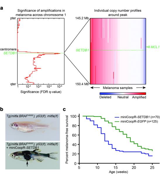
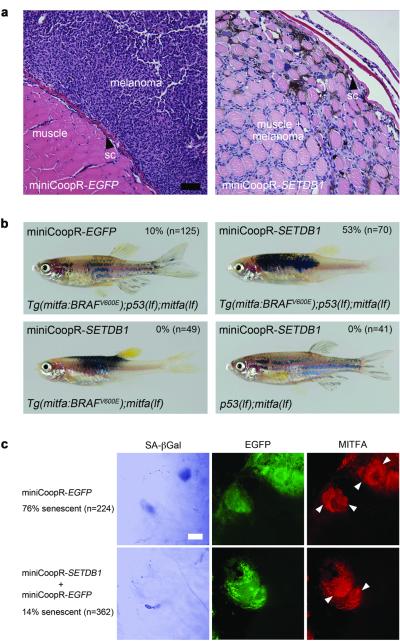
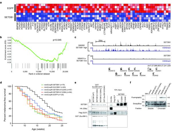
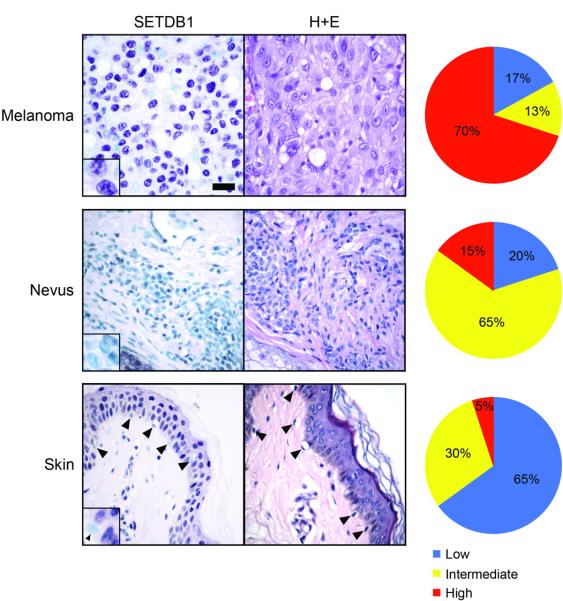
The most common mutation in human melanoma, BRAF(V600E), activates the serine/threonine kinase BRAF and causes excessive activity in the mitogen-activated protein kinase pathway. BRAF(V600E) mutations are also present in benign melanocytic naevi, highlighting the importance of additional genetic alterations in the genesis of malignant tumours. Such changes include recurrent copy number variations that result in the amplification of oncogenes. For certain amplifications, the large number of genes in the interval has precluded an understanding of the cooperating oncogenic events. Here we have used a zebrafish melanoma model to test genes in a recurrently amplified region of chromosome 1 for the ability to cooperate with BRAF(V600E) and accelerate melanoma. SETDB1, an enzyme that methylates histone H3 on lysine 9 (H3K9), was found to accelerate melanoma formation significantly in zebrafish. Chromatin immunoprecipitation coupled with massively parallel DNA sequencing and gene expression analyses uncovered genes, including HOX genes, that are transcriptionally dysregulated in response to increased levels of SETDB1. Our studies establish SETDB1 as an oncogene in melanoma and underscore the role of chromatin factors in regulating tumorigenesis.


Figures

Figure 1
Figure 1. SETDB1accelerates melanoma formation in zebrafish
a, Left, significance of copy number amplification in human melanoma samples determined using genomic identification of significant targets in cancer (GISTIC),. q-values (x-axis) are plotted across chromosome 1. Right, copy number profiles of the 1q21 interval in melanoma samples. The positions of SETDB1 (dotted line) and MCL1 (arrowhead) are indicated. b, The Tg(mitfa:BRAFV600E); p53(lf); mitfa(lf) strain (top) injected with miniCoopR-cloned candidate oncogenes. Animals injected with miniCoopR-SETDB1 (bottom) have rescued melanocytes and rapidly develop melanomas (arrow). c, Melanoma-free survival curve of miniCoopR-SETDB1 (weighted average of 2 independent experiments, n=70) and miniCoopR-EGFP (weighted average of 3 independent experiments, n=125) injected zebrafish.
Figure 2
Figure 2. SETDB1 effects on melanoma cells and melanocytes
a, Hematoxylin and eosin-stained transverse sections of zebrafish melanomas at two weeks post onset. At this time point dorsal miniCoopR-EGFP melanomas (left) display exophytic growth, whereas miniCoopR-SETDB1 melanomas (right) have invaded from the skin, through the collagen-rich stratum compactum of the dermis (sc), into the underlying musculature. Scale bar = 70μm. b, SETDB1 interacts with BRAF(V600E) to affect pigmentation pattern, but a p53(lf) mutation is required to form melanomas. MiniCoopR-EGFP or MiniCoopR-SETDB1 was injected into the indicated transgenic strains. Percentages indicate melanoma incidence at 12 weeks of age. c, SETDB1abrogates BRAF(V600E)-induced senescence. Left, brightfield pseudocolored photomicrographs of SA-βGal staining performed on scale-associated melanocytes. Middle and right, fluorescent photomicrographs of the same melanocytes. miniCoopR-rescued melanocytes in this experiment express mitfa promoter-driven EGFP (middle) and the MITFA protein (right). Multiple nuclei (arrowheads) are present in BRAF(V600E)-expressing melanocytes. Percentage of senescent melanocytes is indicated at left (p = 7.3×10-51, χ2). Scale bar = 10μm.
Figure 3
Figure 3. SETDB1 target gene regulation and HMTase complex formation
a, Heat map of genes downregulated in zebrafish melanomas that overexpress SETDB1 compared with control (EGFP) melanomas. b, Graphical representation of the rank-ordered gene list derived from a panel of human melanoma short-term cultures stratified based on SETDB1 expression level. GSEA shows that homologs of zebrafish SETDB1-downregulated genes are similarly downregulated in human melanomas as levels of SETDB1 increase (ES = -0.35, NES = -1.43, FDR q-val = 0.045, p = 0.045). Arrows indicate positions of HOX genes. c, SETDB1 and H3K9me3 ChIP-Seq profiles at the HOXA locus in human melanoma cells. The number of sequence reads is shown on the y axis. d, Melanoma-free survival curves of zebrafish expressing SUV39H1 (p = 6.74×10⁻⁸ vs. miniCoopR-EGFP, logrank 72) and the methyltransferase-deficient SETDB1(H1224K) (p = 0.24 vs. miniCoopR-SETDB1, p = 8.4×10⁻⁵ vs. miniCoopR-EGFP) and SETDB1(C1226A) (p = 0.20 vs. miniCoopR-SETDB1, p = 1.3×10⁻¹¹ vs. miniCoopR-EGFP) variants. e, In vitro reconstitution of methyltransferase complexes. Sequential purification of GST-tagged SUV39H1, Flag-tagged GLP and HA-tagged G9a proteins was followed by western blotting using antibodies shown on left. f, Histone methylation assays on complexes purified from C2C12 cells.
Figure 4
Figure 4. High expression of SETDB1 protein is common in human melanomas but not nevi or normal melanocytes
Immunohistochemical staining of SETDB1 (left) and hematoxylin and eosin (H+E) staining (center). SETDB1 expression (right) was scored on malignant melanoma (top; n=91), nevi (middle; n=20) and normal skin (bottom; n=20). SETDB1 expression was measured as described in Methods. Summarized and raw data from two independent antibodies are described in Supplementary Tables 4 and 5, respectively. Scale bar = 30μm, insets are 2.5x magnified.

References

    1. Davies H, et al. Mutations of the BRAF gene in human cancer. Nature. 2002;417(6892):949–954. - PubMed
    1. Wan PT, et al. Mechanism of activation of the RAF-ERK signaling pathway by oncogenic mutations of B-RAF. Cell. 2004;116(6):855–867. - PubMed
    1. Pollock PM, et al. High frequency of BRAF mutations in nevi. Nat Genet. 2003;33(1):19–20. - PubMed
    1. Curtin JA, et al. Distinct sets of genetic alterations in melanoma. N Engl J Med. 2005;353(20):2135–2147. - PubMed
    1. Garraway LA, et al. Integrative genomic analyses identify MITF as a lineage survival oncogene amplified in malignant melanoma. Nature. 2005;436(7047):117–122. - PubMed

Publication types

MeSH terms

Substances

Associated data