

Neurologic complication following spinal epidural anesthesia in a patient with spinal intradural extramedullary tumor
- PMID: 21430985
- PMCID: PMC3053553
- DOI: 10.3340/jkns.2010.48.6.544
Neurologic complication following spinal epidural anesthesia in a patient with spinal intradural extramedullary tumor
Abstract
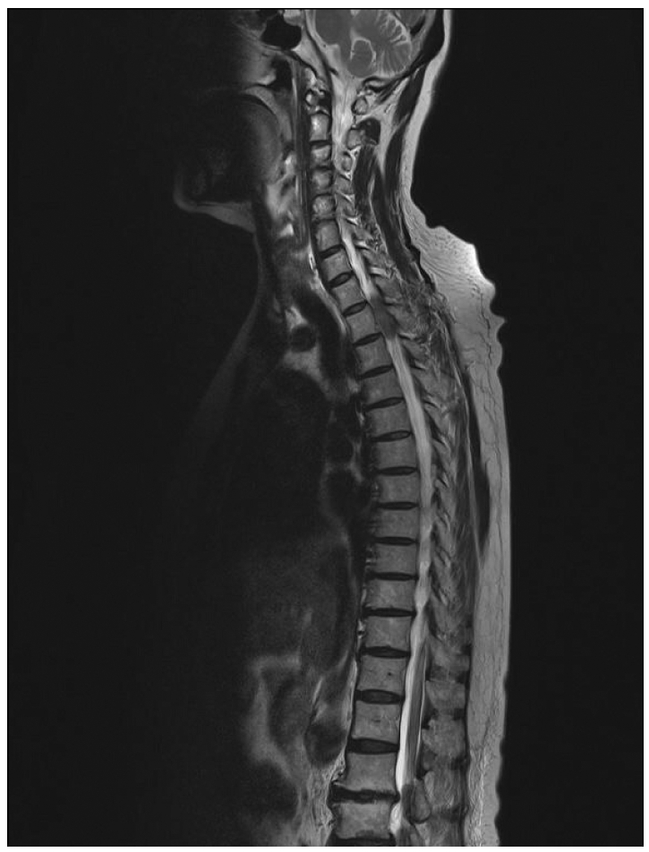
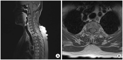
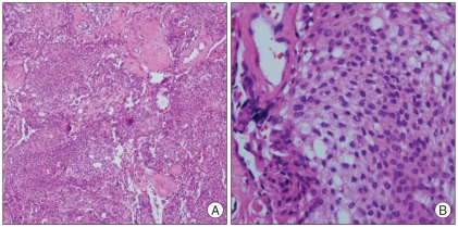
Paraplegia following spinal epidural anesthesia is extremely rare. Various lesions for neurologic complications have been documented in the literature. We report a 66-year-old female who developed paraplegia after left knee surgery for osteoarthritis under spinal epidural anesthesia. In the recovery room, paraplegia and numbness below T4 vertebra was checked. A magnetic resonance image (MRI) scan showed a spinal thoracic intradural extramedullary (IDEM) tumor. After extirpation of the tumor, the motor weakness improved to the grade of 3/5. If a neurologic deficit following spinal epidural anesthesia does not resolve, a MRI should be performed without delay to accurately diagnose the cause of the deficit and optimal treatment should be rendered for the causative lesion.
Keywords: Spinal epidural anesthesia; Spinal intradural extramedullary tumor.
Figures
Similar articles
-
A rare case of intradural and extramedullary epidermoid cyst after repetitive epidural anesthesia: case report and review of the literature.World J Surg Oncol. 2017 Jul 17;15(1):131. doi: 10.1186/s12957-017-1186-4. World J Surg Oncol. 2017. PMID: 28716031 Free PMC article. Review.
-
Paraplegia caused by giant intradural herniation of a lumbar disk after combined spinal-epidural anesthesia in total hip arthroplasty.J Clin Anesth. 2016 Aug;32:169-71. doi: 10.1016/j.jclinane.2016.02.025. Epub 2016 Apr 20. J Clin Anesth. 2016. PMID: 27290969
-
CT and MRI presentation of intradural epidural angiolipoma of the thoracic spinal canal (with a case report).Radiol Case Rep. 2023 Feb 27;18(5):1721-1726. doi: 10.1016/j.radcr.2023.01.095. eCollection 2023 May. Radiol Case Rep. 2023. PMID: 36895895 Free PMC article.
-
Spinal intradural, extramedullary anaplastic ependymoma with an extradural component: Case report and review of the literature.Surg Neurol Int. 2011;2:119. doi: 10.4103/2152-7806.84246. Epub 2011 Aug 30. Surg Neurol Int. 2011. PMID: 21918734 Free PMC article.
-
Primary intradural extramedullary spinal mesenchymal chondrosarcoma: case report and literature review.BMC Musculoskelet Disord. 2019 Sep 4;20(1):408. doi: 10.1186/s12891-019-2799-2. BMC Musculoskelet Disord. 2019. PMID: 31484514 Free PMC article. Review.
Cited by
-
Paresthesia and sensory deficits on the unilateral leg arising from an unrecognized intramedullary tumor after spinal anesthesia.Korean J Anesthesiol. 2013 May;64(5):472-3. doi: 10.4097/kjae.2013.64.5.472. Korean J Anesthesiol. 2013. PMID: 23741575 Free PMC article. No abstract available.
-
Cauda Equina Syndrome Secondary to Incidental Hemorrhagic Ependymoma Following Spinal Anesthesia: A Case Report and Comprehensive Review.Cureus. 2023 Jun 15;15(6):e40490. doi: 10.7759/cureus.40490. eCollection 2023 Jun. Cureus. 2023. PMID: 37333038 Free PMC article.
-
Neurological complications following spinal anaesthesia in a patient with congenital absence of lumbar vertebra.Indian J Anaesth. 2014 Jul;58(4):484-6. doi: 10.4103/0019-5049.139021. Indian J Anaesth. 2014. PMID: 25197127 Free PMC article. No abstract available.
-
Spinal coning after lumbar puncture in a patient with undiagnosed giant cervical neurofibroma.Ann Indian Acad Neurol. 2013 Jul;16(3):440-2. doi: 10.4103/0972-2327.116935. Ann Indian Acad Neurol. 2013. PMID: 24101840 Free PMC article.
-
Treatment results in the differential surgery of intradural extramedullary schwannoma of 110 cases.PLoS One. 2013 May 27;8(5):e63867. doi: 10.1371/journal.pone.0063867. Print 2013. PLoS One. 2013. PMID: 23724010 Free PMC article.
References
-
- Cherng YG, Chen IY, Liu FL, Wang MH. Paraplegia following spinal anesthesia in a patient with an undiagnosed metastatic spinal tumor. Acta Anaesthesiol Taiwan. 2008;46:86–90. - PubMed
-
- Faccenda KA, Finucane BT. Complications of regional anesthesia. Drug Saf. 2001;24:413–442. - PubMed
-
- Greene NM. Neurological sequelae of spinal anesthesia. Anesthesiology. 1961;22:682–698. - PubMed
-
- Hollis PH, MAlis LI, Zappulla RA. Neurological deterioration after lumbar puncture below complete spinal subarachnoid block. J Neurosurg. 1986;64:253–256. - PubMed
-
- Horlocker TT, Abel MD, Messick JM, Jr, Schroeder DR. Small risk of serious neurologic complications related to lumbar epidural catheter placement in anesthetized patients. Anesth Analg. 2003;96:1547–1552. - PubMed
Publication types
LinkOut - more resources
Full Text Sources
