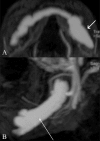
Case report: Combined transarterial and direct approaches for embolization of a large mandibular arteriovenous malformation
- PMID: 21431023
- PMCID: PMC3056373
- DOI: 10.4103/0971-3026.76044
Case report: Combined transarterial and direct approaches for embolization of a large mandibular arteriovenous malformation
Abstract
Arteriovenous malformations (AVMs) that involve the mandible are difficult lesions to treat, with traditional options being surgery and embolization. This article describes a large mandibular AVM that was treated with embolization using transarterial as well as direct puncture approaches. Follow-up imaging showed thrombosis of the vascular spaces of the malformation. There were no complications. The patient is doing well and is on follow-up.
Keywords: Arteriovenous malformation; embolization; mandible.
Conflict of interest statement
Figures


Similar articles
-
Retrograde transvenous balloon-assisted Onyx embolization of mandibular arteriovenous malformation after hemorrhage.Radiol Case Rep. 2018 Dec 13;14(3):348-353. doi: 10.1016/j.radcr.2018.11.016. eCollection 2019 Mar. Radiol Case Rep. 2018. PMID: 30581522 Free PMC article.
-
Osseous regeneration after embolization of mandibular arteriovenous malformation.Chang Gung Med J. 2003 Dec;26(12):937-42. Chang Gung Med J. 2003. PMID: 15008331
-
Treatment of mandibular arteriovenous malformation by transvenous embolization: A case report.Head Neck. 1999 Sep;21(6):574-7. doi: 10.1002/(sici)1097-0347(199909)21:6<574::aid-hed12>3.0.co;2-d. Head Neck. 1999. PMID: 10449675
-
Curative Embolization of Arteriovenous Malformations.World Neurosurg. 2019 Sep;129:467-486. doi: 10.1016/j.wneu.2019.01.166. Epub 2019 Feb 5. World Neurosurg. 2019. PMID: 30735875 Review.
-
How to Treat Peripheral Arteriovenous Malformations.Korean J Radiol. 2021 Apr;22(4):568-576. doi: 10.3348/kjr.2020.0981. Epub 2020 Dec 21. Korean J Radiol. 2021. PMID: 33543847 Free PMC article. Review.
Cited by
-
Endovascular embolisation of a complex mandibular AVM: a hybrid transarterial and transvenous approach.BMJ Case Rep. 2023 Jan 24;16(1):e251589. doi: 10.1136/bcr-2022-251589. BMJ Case Rep. 2023. PMID: 36693701 Free PMC article.
References
-
- Chen W, Wang J, Li J, Xu L. Comprehensive treatment of arteriovenous malformations in the oral and maxillofacial region. J Oral Maxillofac Surg. 2005;63:1484–8. - PubMed
-
- Motamedi MH, Behnia H, Motamedi MR. Surgical technique for the treatment of high-flow arteriovenous malformations of the mandible. J Craniomaxillofac Surg. 2000;28:238–42. - PubMed
-
- Beek FJ, ten Broek FW, van Schaik JP, Mali WP. Transvenous embolisation of an arteriovenous malformation of the mandible via a femoral approach. Pediatr Radiol. 1997;27:855–7. - PubMed
-
- Guibert-Tranier F, Piton J, Riche MC, Merland JJ, Caille JM. Vascular malformations of the mandible (intraosseous haemangiomas). The importance of preoperative embolization. A study of 9 cases. Eur J Radiol. 1982;2:257–72. - PubMed
Publication types
LinkOut - more resources
Full Text Sources

