Cross-sectional area of posterior extensor muscles of the cervical spine in asymptomatic subjects: a 10-year longitudinal magnetic resonance imaging study
- PMID: 21431426
- PMCID: PMC3175907
- DOI: 10.1007/s00586-011-1774-x
Cross-sectional area of posterior extensor muscles of the cervical spine in asymptomatic subjects: a 10-year longitudinal magnetic resonance imaging study
Abstract
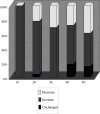
There has been no prospective study on age-related changes of the extensor muscles of the cervical spine in healthy subjects. This study was conducted to elucidate any association between the changes in cross-sectional area of the extensor muscles of the cervical spine on MRIs and cervical disc degeneration or the development of clinical symptoms. Sixty-two subjects who underwent MR imaging by a 1.5-Tesla machine between 1993 and 1996 as asymptomatic volunteers in a previous study were recruited again 10 years later for this follow-up study. The mean interval between the studies was 11.0 ± 0.7 years. The cross-sectional areas of the multifidus, semispinalis cervicis, semispinalis capitis, and splenius capitis at C3-C4, C4-C5, and C5-C6 intervertebral levels were measured on T2-weighted axial images using Image J 1.42. The mean cross-sectional areas of the deep extensor muscles were 1,396.8 ± 337.6 mm(2) at the C3-C4 level, 1,514.7 ± 381.0 mm(2) at the C4-C5 level, and 1,542.8 ± 373.5 mm(2) at the C5-C6 level in the previous investigation. The cross-sectional areas were 1,498.7 ± 374.4 mm(2) at the C3-C4 level, 1,569.9 ± 390.9 mm(2) at the C4-C5 level, and 1,599.6 ± 364.3 mm(2) at the 10-year follow-up. An increase in the cross-sectional area of the muscles was more frequently observed in subjects in their tens to thirties in the initial study, while a decrease was more frequently observed in those in their forties and older in the initial study. Disc degeneration was not correlated with a change in extensor muscle volume. Development of shoulder stiffness during follow-up was significantly negatively correlated with a change in the cross-sectional area of the deep extensor muscles.
Figures


Similar articles
-
Cross-sectional area of the posterior extensor muscles of the cervical spine in whiplash injury patients versus healthy volunteers--10 year follow-up MR study.Injury. 2012 Jun;43(6):912-6. doi: 10.1016/j.injury.2012.01.017. Epub 2012 Feb 4. Injury. 2012. PMID: 22310029
-
Longitudinal MRI study over 20 years of cervical posterior extensor muscle area in asymptomatic subjects.Sci Rep. 2025 Jul 1;15(1):21826. doi: 10.1038/s41598-025-08055-6. Sci Rep. 2025. PMID: 40595211 Free PMC article.
-
Magnetic resonance imaging study of cross-sectional area of the cervical extensor musculature in an asymptomatic cohort.Clin Anat. 2007 Jan;20(1):35-40. doi: 10.1002/ca.20252. Clin Anat. 2007. PMID: 16302247
-
Function and structure of the deep cervical extensor muscles in patients with neck pain.Man Ther. 2013 Oct;18(5):360-6. doi: 10.1016/j.math.2013.05.009. Epub 2013 Jul 12. Man Ther. 2013. PMID: 23849933 Review.
-
Normalization technique to build patient specific muscle model in finite element head neck spine.Med Eng Phys. 2022 Sep;107:103857. doi: 10.1016/j.medengphy.2022.103857. Epub 2022 Jul 21. Med Eng Phys. 2022. PMID: 36068040 Review.
Cited by
-
Evaluation of ACL mid-substance cross-sectional area for reconstructed autograft selection.Knee Surg Sports Traumatol Arthrosc. 2014 Jan;22(1):207-13. doi: 10.1007/s00167-012-2356-0. Epub 2012 Dec 22. Knee Surg Sports Traumatol Arthrosc. 2014. PMID: 23263230
-
Magnetic resonance imaging-based relationships between neck muscle cross-sectional area and neck circumference for adults and children.Eur Spine J. 2013 Feb;22(2):446-52. doi: 10.1007/s00586-012-2482-x. Epub 2012 Aug 28. Eur Spine J. 2013. PMID: 22926433 Free PMC article.
-
Clinical and Radiographic Outcomes of Modified Posterior Atlantoaxial Temporary Fixation with Preservation of Semispinalis Cervicis: A Comparative Study.Global Spine J. 2024 Jan;14(1):272-282. doi: 10.1177/21925682221103832. Epub 2022 May 22. Global Spine J. 2024. PMID: 35603736 Free PMC article.
-
Dropped Head Syndrome Treated with Physical Therapy Based on the Concept of Athletic Rehabilitation.Case Rep Orthop. 2020 Dec 8;2020:8811148. doi: 10.1155/2020/8811148. eCollection 2020. Case Rep Orthop. 2020. PMID: 33489397 Free PMC article.
-
Inter-rater reliability of trunk muscle morphometric analysis.J Back Musculoskelet Rehabil. 2015;28(1):181-90. doi: 10.3233/BMR-140552. J Back Musculoskelet Rehabil. 2015. PMID: 25628042 Free PMC article.
References
-
- Booth FW, Weeden SH, Tseng BS. Effect of aging on human skeletal muscle and motor function. Med Sci Sports Exerc. 1994;26:556–560. - PubMed
-
- Cohn SH, Vartsky D, Yasumura S, et al. Compartmental body composition based on total-body nitrogen, potassium, and calcium. Am J Physiol. 1980;239:524–530. - PubMed
Publication types
MeSH terms
LinkOut - more resources
Full Text Sources
Medical
Miscellaneous

