

Morphine potentiates neuropathogenesis of SIV infection in rhesus macaques
- PMID: 21431470
- PMCID: PMC3270698
- DOI: 10.1007/s11481-011-9272-9
Morphine potentiates neuropathogenesis of SIV infection in rhesus macaques
Abstract
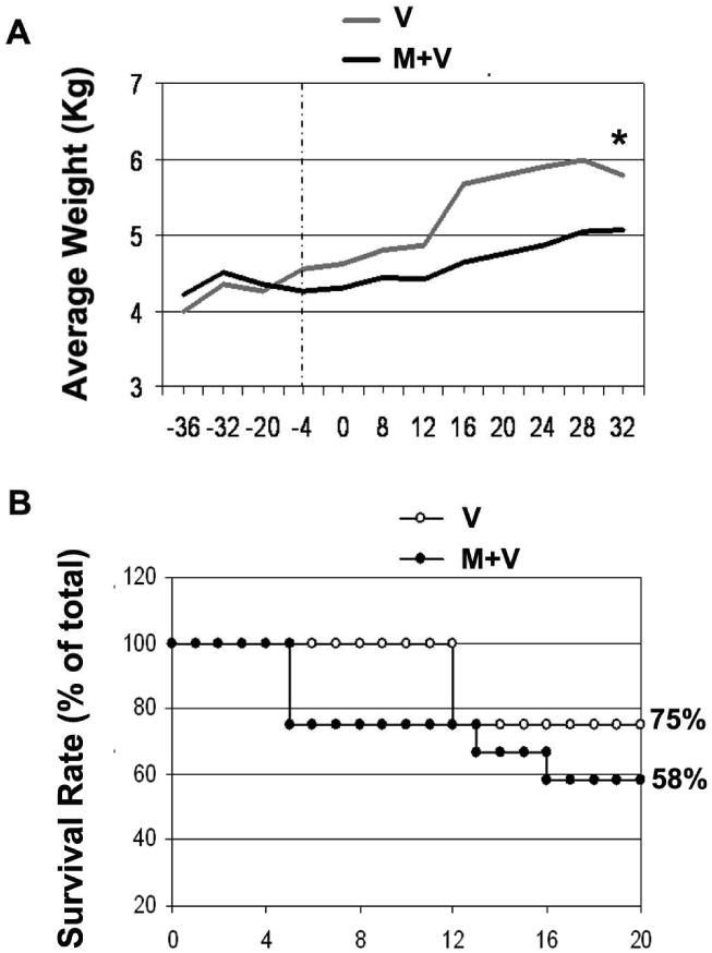
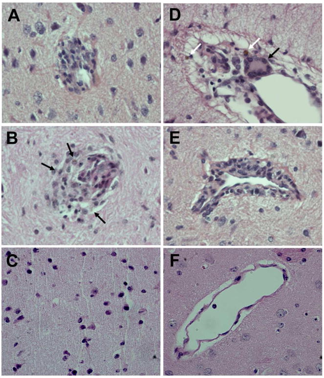
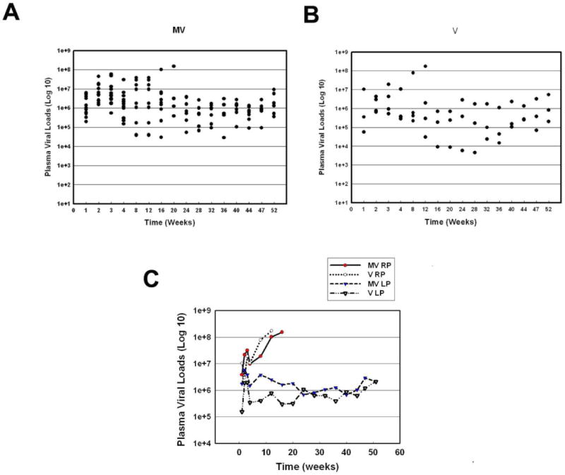
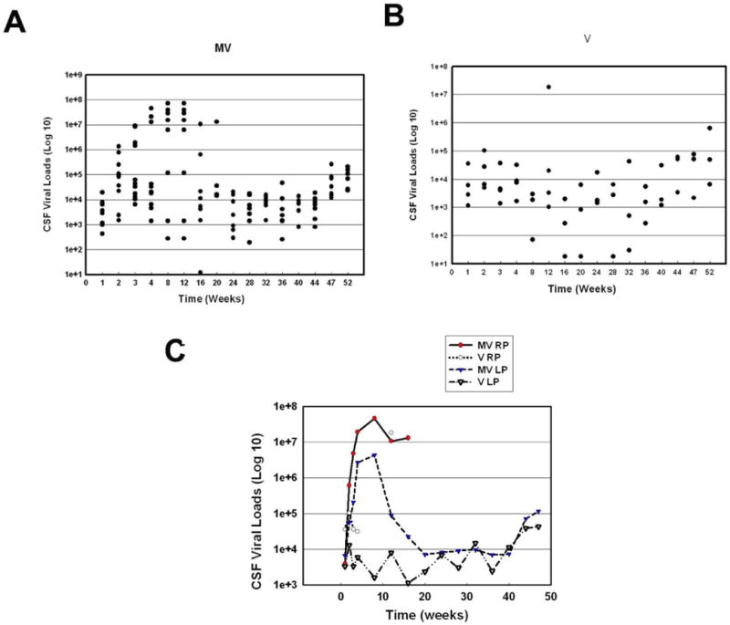
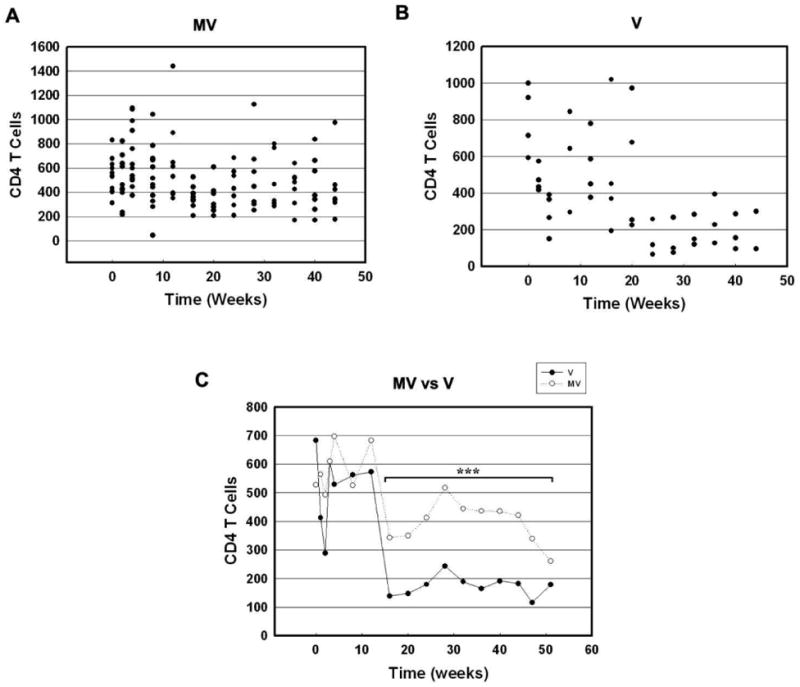
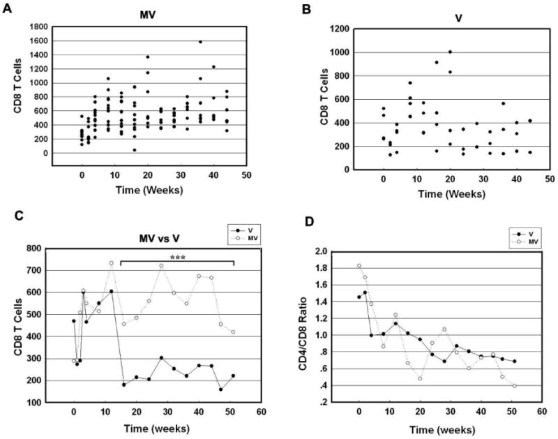
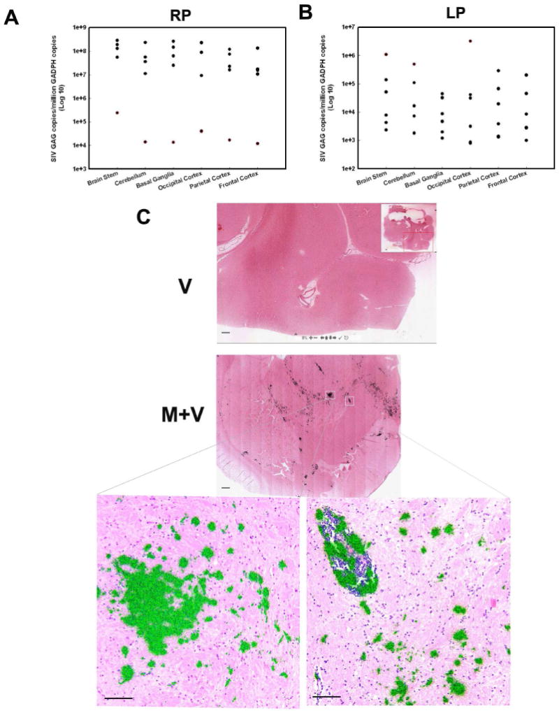
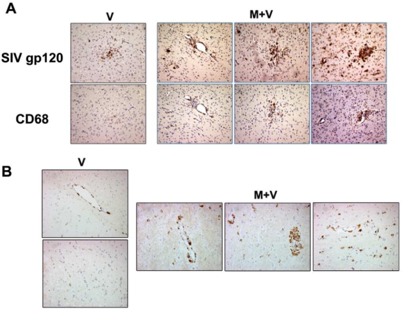
Despite the advent of antiretroviral therapy, complications of HIV-1 infection with concurrent drug abuse are an emerging problem. Opiates are well known to modulate immune responses by preventing the development of cell-mediated immune responses. Their effect on the pathogenesis of HIV-1 infection however remains controversial. Using the simian immunodeficiency virus/macaque model of HIV pathogenesis, we sought to explore the impact of morphine on disease progression and pathogenesis. Sixteen rhesus macaques were divided into two groups; four were administered saline and 12 others morphine routinely. Both groups of animals were then inoculated with SIVmacR71/17E and followed longitudinally for disease pathogenesis. The morphine group (M+V) exhibited a trend towards higher mortality rates and retardation in weight gain compared to the virus-alone group. Interestingly, a subset of M+V animals succumbed to disease within weeks post-infection. These rapid progressors also exhibited a higher incidence of other end-organ pathologies. Despite the higher numbers of circulating CD4+ and CD8+ T cells in the M+V group, CD4/CD8 ratios between the groups remained unchanged. Plasma and CSF viral load in the M+V group was at least a log higher than the control group. Similarly, there was a trend toward increased virus build-up in the brains of M+V animals compared with controls. A novel finding of this study was the increased influx of infected monocyte/macrophages in the brains of M+V animals.
Figures
Similar articles
-
Effect of morphine on the neuropathogenesis of SIVmac infection in Indian Rhesus Macaques.J Neuroimmune Pharmacol. 2008 Mar;3(1):12-25. doi: 10.1007/s11481-007-9085-z. Epub 2007 Sep 13. J Neuroimmune Pharmacol. 2008. PMID: 18247128
-
Critical Role for Monocytes/Macrophages in Rapid Progression to AIDS in Pediatric Simian Immunodeficiency Virus-Infected Rhesus Macaques.J Virol. 2017 Aug 10;91(17):e00379-17. doi: 10.1128/JVI.00379-17. Print 2017 Sep 1. J Virol. 2017. PMID: 28566378 Free PMC article.
-
Elite Control, Gut CD4 T Cell Sparing, and Enhanced Mucosal T Cell Responses in Macaca nemestrina Infected by a Simian Immunodeficiency Virus Lacking a gp41 Trafficking Motif.J Virol. 2015 Oct;89(20):10156-75. doi: 10.1128/JVI.01134-15. Epub 2015 Jul 29. J Virol. 2015. PMID: 26223646 Free PMC article.
-
Chronic morphine exposure causes pronounced virus replication in cerebral compartment and accelerated onset of AIDS in SIV/SHIV-infected Indian rhesus macaques.Virology. 2006 Oct 10;354(1):192-206. doi: 10.1016/j.virol.2006.06.020. Epub 2006 Jul 28. Virology. 2006. PMID: 16876224
-
Simian Immunodeficiency Virus-Infected Memory CD4+ T Cells Infiltrate to the Site of Infected Macrophages in the Neuroparenchyma of a Chronic Macaque Model of Neurological Complications of AIDS.mBio. 2020 Apr 21;11(2):e00602-20. doi: 10.1128/mBio.00602-20. mBio. 2020. PMID: 32317323 Free PMC article.
Cited by
-
Productive infection of human neural progenitor cells by R5 tropic HIV-1: opiate co-exposure heightens infectivity and functional vulnerability.AIDS. 2017 Mar 27;31(6):753-764. doi: 10.1097/QAD.0000000000001398. AIDS. 2017. PMID: 28099189 Free PMC article.
-
Opioids and Opioid Maintenance Therapies: Their Impact on Monocyte-Mediated HIV Neuropathogenesis.Curr HIV Res. 2016;14(5):417-430. doi: 10.2174/1570162x14666160324124132. Curr HIV Res. 2016. PMID: 27009099 Free PMC article. Review.
-
Morphine produces immunosuppressive effects in nonhuman primates at the proteomic and cellular levels.Mol Cell Proteomics. 2012 Sep;11(9):605-18. doi: 10.1074/mcp.M111.016121. Epub 2012 May 11. Mol Cell Proteomics. 2012. PMID: 22580588 Free PMC article.
-
Cell-type specific differences in antiretroviral penetration and the effects of HIV-1 Tat and morphine among primary human brain endothelial cells, astrocytes, pericytes, and microglia.Neurosci Lett. 2019 Nov 1;712:134475. doi: 10.1016/j.neulet.2019.134475. Epub 2019 Sep 3. Neurosci Lett. 2019. PMID: 31491466 Free PMC article.
-
HIV-1 Tat and morphine interactions dynamically shift striatal monoamine levels and exploratory behaviors over time.J Neurochem. 2024 Mar;168(3):185-204. doi: 10.1111/jnc.16057. Epub 2024 Feb 3. J Neurochem. 2024. PMID: 38308495 Free PMC article.
References
-
- Bell JE, Donaldson YK, Lowrie S, McKenzie CA, Elton RA, Chiswick A, Brettle RP, Ironside JW, Simmonds P. Influence of risk group and zidovudine therapy on the development of HIV encephalitis and cognitive impairment in AIDS patients. AIDS. 1996;10:493–499. - PubMed
-
- Bouwman FH, Skolasky RL, Hes D, Selnes OA, Glass JD, Nance-Sproson TE, Royal W, Dal Pan GJ, McArthur JC. Variable progression of HIV-associated dementia. Neurology. 1998;50:1814–1820. - PubMed
Publication types
MeSH terms
Substances
Grants and funding
LinkOut - more resources
Full Text Sources
Research Materials
