

Correlations between ventricular enlargement and gray and white matter volumes of cortex, thalamus, striatum, and internal capsule in schizophrenia
- PMID: 21431919
- PMCID: PMC3182327
- DOI: 10.1007/s00406-011-0202-x
Correlations between ventricular enlargement and gray and white matter volumes of cortex, thalamus, striatum, and internal capsule in schizophrenia
Abstract
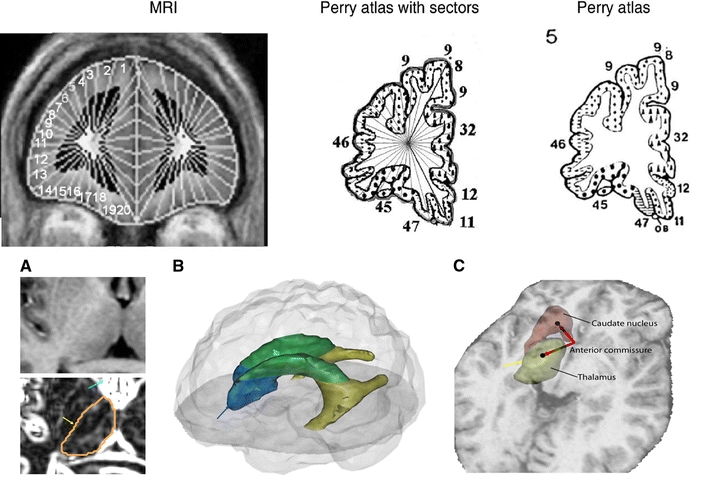
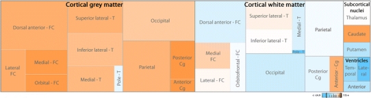
Ventricular enlargement is one of the most consistent abnormal structural brain findings in schizophrenia and has been used to infer brain shrinkage. However, whether ventricular enlargement is related to local overlying cortex and/or adjacent subcortical structures or whether it is related to brain volume change globally has not been assessed. We systematically assessed interrelations of ventricular volumes with gray and white matter volumes of 40 Brodmann areas (BAs), the thalamus and its medial dorsal nucleus and pulvinar, the internal capsule, caudate and putamen. We acquired structural MRI ( patients with schizophrenia (n = 64) and healthy controls (n = 56)) and diffusion tensor fractional anisotropy (FA) (untreated schizophrenia n = 19, controls n = 32). Volumes were assessed by manual tracing of central structures and a semi-automated parcellation of BAs. Patients with schizophrenia had increased ventricular size associated with decreased cortical gray matter volumes widely across the brain; a similar but less pronounced pattern was seen in normal controls; local correlations (e.g. temporal horn with temporal lobe volume) were not appreciably higher than non-local correlations (e.g. temporal horn with prefrontal volume). White matter regions adjacent to the ventricles similarly did not reveal strong regional relationships. FA and center of mass of the anterior limb of the internal capsule also appeared differentially influenced by ventricular volume but findings were similarly not regional. Taken together, these findings indicate that ventricular enlargement is globally interrelated with gray matter volume diminution but not directly correlated with volume loss in the immediately adjacent caudate, putamen, or internal capsule.
Figures
Similar articles
-
Cerebral grey, white matter and csf in never-medicated, first-episode schizophrenia.Schizophr Res. 2007 Jan;89(1-3):12-21. doi: 10.1016/j.schres.2006.09.009. Epub 2006 Nov 13. Schizophr Res. 2007. PMID: 17098398
-
Focal white matter density changes in schizophrenia: reduced inter-hemispheric connectivity.Neuroimage. 2004 Jan;21(1):27-35. doi: 10.1016/j.neuroimage.2003.09.026. Neuroimage. 2004. PMID: 14741639
-
Altered white matter/gray matter proportions in the striatum of patients with schizophrenia: a volumetric MRI study.Am J Psychiatry. 2005 Dec;162(12):2315-21. doi: 10.1176/appi.ajp.162.12.2315. Am J Psychiatry. 2005. PMID: 16330596
-
[Connectivity analyses of white matters in schizophrenia].Seishin Shinkeigaku Zasshi. 2013;115(8):880-6. Seishin Shinkeigaku Zasshi. 2013. PMID: 24167969 Review. Japanese.
-
A review of MRI findings in schizophrenia.Schizophr Res. 2001 Apr 15;49(1-2):1-52. doi: 10.1016/s0920-9964(01)00163-3. Schizophr Res. 2001. PMID: 11343862 Free PMC article. Review.
Cited by
-
Cerebral Folate Metabolism in Post-Mortem Alzheimer's Disease Tissues: A Small Cohort Study.Int J Mol Sci. 2022 Dec 30;24(1):660. doi: 10.3390/ijms24010660. Int J Mol Sci. 2022. PMID: 36614107 Free PMC article.
-
Subcortical Brain Volume Abnormalities in Individuals With an At-risk Mental State.Schizophr Bull. 2020 Jul 8;46(4):834-845. doi: 10.1093/schbul/sbaa011. Schizophr Bull. 2020. PMID: 32162659 Free PMC article.
-
From generation of biomarkers to treatment and psychosocial aspects of psychosis.Eur Arch Psychiatry Clin Neurosci. 2011 Oct;261(7):457-8. doi: 10.1007/s00406-011-0262-y. Eur Arch Psychiatry Clin Neurosci. 2011. PMID: 21927833 Free PMC article. No abstract available.
-
Gray matter volume drives the brain age gap in schizophrenia: a SHAP study.Schizophrenia (Heidelb). 2023 Jan 9;9(1):3. doi: 10.1038/s41537-022-00330-z. Schizophrenia (Heidelb). 2023. PMID: 36624107 Free PMC article.
-
Four-way multimodal fusion of 7 T imaging data using an mCCA+jICA model in first-episode schizophrenia.Hum Brain Mapp. 2018 Apr;39(4):1475-1488. doi: 10.1002/hbm.23906. Epub 2018 Jan 9. Hum Brain Mapp. 2018. PMID: 29315951 Free PMC article.
References
-
- Wright IC, et al. Meta-analysis of regional brain volumes in schizophrenia. Am J Psychiatry. 2000;157(1):16–25. - PubMed
MeSH terms
LinkOut - more resources
Full Text Sources
Medical
