. 2011 May;224(1):67-77.
doi: 10.1002/path.2851. Epub 2011 Mar 22.

Prominent expression of sialyl Lewis X-capped core 2-branched O-glycans on high endothelial venule-like vessels in gastric MALT lymphoma

Affiliations

Prominent expression of sialyl Lewis X-capped core 2-branched O-glycans on high endothelial venule-like vessels in gastric MALT lymphoma

Motohiro Kobayashi et al. J Pathol. 2011 May.

Abstract

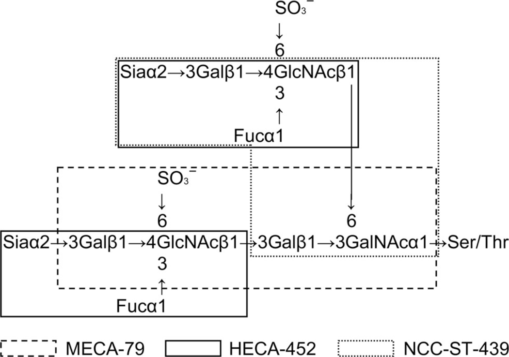
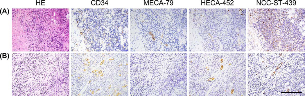
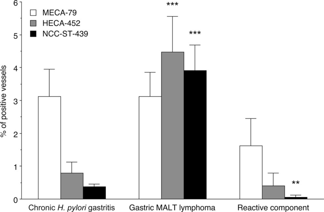
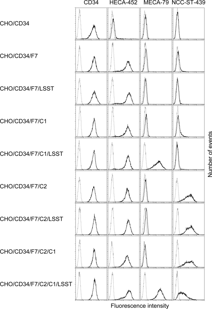
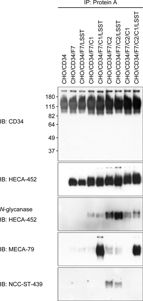
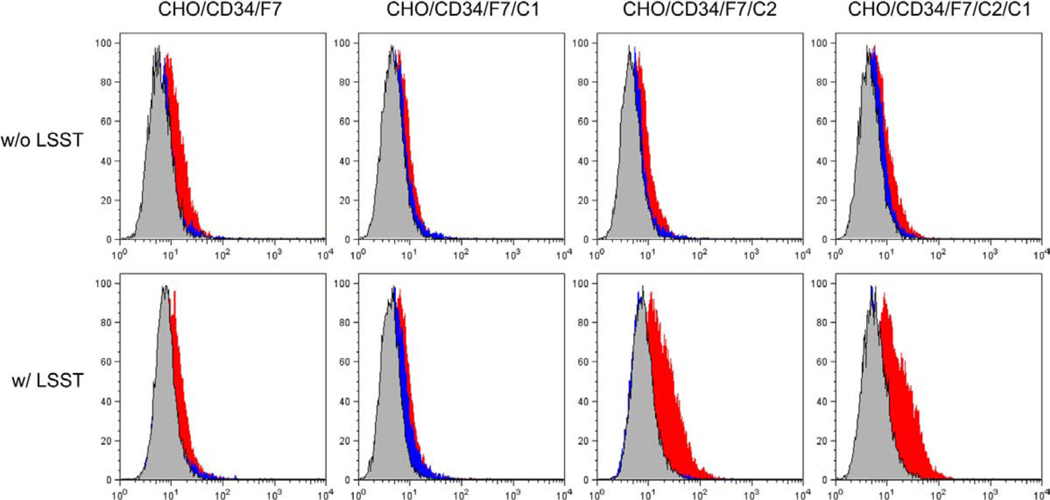
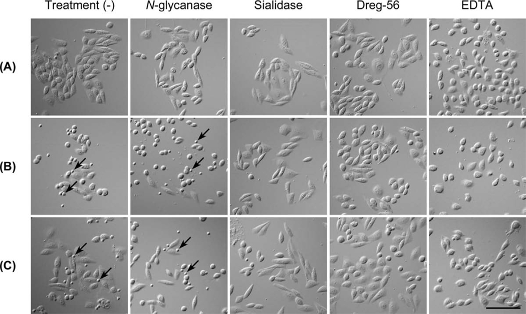
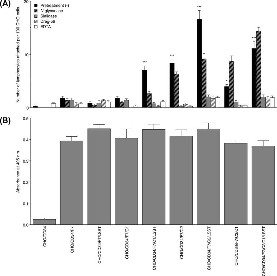
High endothelial venule (HEV)-like vessels have been observed in gastric B cell lymphoma of mucosa-associated lymphoid tissue type (MALT lymphoma), as well as in its preceding lesion, chronic Helicobacter pylori gastritis. Previously we reported that glycans on HEV-like vessels in the latter lesion served as L-selectin ligands, although their function is unclear. We have investigated sialyl Lewis X (sLeX)-related glycoepitopes and found that MECA-79(-) /HECA-452(+) /NCC-ST-439(+) HEV-like vessels preferentially mark gastric MALT lymphoma compared to chronic H. pylori gastritis. We then constructed CHO cell lines expressing potential MECA-79(-) /HECA-452(+) /NCC-ST-439(+) glycans, as well as other sLeX-type glycans, on CD34 and evaluated L-selectin binding to those cells, using L-selectin-IgM chimera binding and lymphocyte adhesion assays. L-selectin-IgM chimeras bound to CHO cells expressing 6-sulpho-sLeX attached to core 2-branched O-glycans with or without 6-sulpho-sLeX attached to extended core 1 O-glycans, but only marginally to other CHO cell lines. By contrast, CHO cells expressing 6-sulpho-sLeX attached to extended core 1 and/or core 2-branched O-glycans, as well as non-sulphated sLeX attached to core 2-branched O-glycans, showed substantial lymphocyte binding, while binding was negligible on lines expressing 6-sulpho- and non-sulphated sLeX attached to N-glycans and non-sulphated sLeX attached to extended core 1 O-glycans. These results indicate that MECA-79(-) /HECA-452(+) /NCC-ST-439(+) glycans, specifically, 6-sulpho- and non-sulphated sLeXs attached to core 2-branched O-glycans, expressed on HEV-like vessels in gastric MALT lymphoma function as L-selectin ligands and likely contribute to H. pylori-specific T cell recruitment in the progression of gastric MALT lymphoma.


Conflict of interest statement

No conflicts of interest were declared.

Figures

Figure 1
Figure 1
Schematic representation of carbohydrate structure of L-selectin ligands. 6-sulfo sLeX attached to extended core 1 and/or core 2-branched O-glycans functions as an L-selectin ligand. Epitopes for monoclonal antibodies MECA-79 (6-sulfo N-acetyllactosamine attached to extended core 1 O-glycans), HECA-452 (sLeX, regardless of GlcNAc-6-O-sulfation), and NCC-ST-439 (sLeX attached to core 2-branched O-glycans, regardless of GlcNAc-6-O-sulfation) are shown.
Figure 2
Figure 2
HEV-like vessels appear in chronic H. pylori gastritis (A) and gastric MALT lymphoma (B). Serial tissue sections were stained with hematoxylin and eosin (HE), and immunostained for CD34 as a marker of vascular endothelial cells, MECA-79, HECA-452, and NCC-ST-439. In chronic H. pylori gastritis, more than half of MECA-79+ vessels are HECA-452/NCC-ST-439. Conversely, in gastric MALT lymphoma, MECA-79 but HECA-452+/NCC-ST-439+ vessels are frequently observed. Bar = 200 µm.
Figure 3
Figure 3
Frequency of MECA-79+, HECA-452+, and NCC-ST-439+ HEV-like vessels in chronic H. pylori gastritis (left), gastric MALT lymphoma (middle), and its accompanied reactive component (right). In chronic H. pylori gastritis, the percentage of MECA-79+ vessels is greater than that of HECA-452+ and NCC-ST-439+ vessels, with high statistical significance (p < 0.001). Conversely, in gastric MALT lymphoma, the percentages of MECA-79+, HECA-452+, and NCC-ST-439+ vessels do not differ significantly. The percentage of HECA-452+ and NCC-ST-439+ but not MECA-79+ vessels in gastric MALT lymphoma is greater than that seen in chronic H. pylori gastritis, with statistical significance. In addition, the percentages of MECA-79+, HECA-452+, and NCC-ST-439+ vessels in accompanied reactive component of gastric MALT lymphoma shows similar patterns seen in chronic H. pylori gastritis. Data are presented as means ± SEM (n = 31 in chronic H. pylori gastritis, n = 22 in gastric MALT lymphoma, n = 3 in accompanied reactive component of gastric MALT lymphoma). ***, p < 0.001; **, p < 0.01.
Figure 4
Figure 4
FACS analysis of CHO cell lines expressing various sugar chains. Each CHO line described in Table 1 was tested for CD34, HECA-452, MECA-79, and NCC-ST-439 reactivities. Lighter gray lines represent negative control resulting from omitting the primary antibody.
Figure 5
Figure 5
Expression of a particular sLeX-type glycans on CD34 obtained from CHO cell lines described in Table 1. CD34 obtained from all lines except control CHO/CD34 cells are positive for HECA-452, and such HECA-452 glycoepitopes are eliminated after treatment with N-glycanase. The MECA-79 epitope is detected in CHO cells transfected with both Core1-β3GlcNAcT and LSST (GlcNAc6ST-2). The NCC-ST-439 epitope is detected in CHO/CD34/F7/C2 with or without transfection with LSST.
Figure 6
Figure 6
L-selectin•IgM chimera binding assay of CHO cell lines. CHO cell lines listed in Table 1 were tested. The L-selectin•IgM chimera most robustly binds to the CHO/CD34/F7/C2/LSST and CHO/CD34/F7/C2/C1/LSST lines at almost equivalent levels. The remaining lines show only marginal L-selectin•IgM binding. Red histograms represent L-selectin•IgM chimera bindings. Blue and gray histograms indicate control experiments using cells treated with sialidase and EDTA, respectively.
Figure 7
Figure 7
Lymphocyte adhesion to CHO cell lines expressing various sugar chains. CHO lines listed in Table 1 were examined. Lymphocyte adhesion to CHO/CD34 (A), CHO/CD34/F7/C2 (B), and CHO/CD34/F7/C2/LSST (C) observed with Nomarski differential interference optics is shown. Arrows indicate adhered lymphocytes. Note that treatments with sialidase, Dreg-56, and EDTA completely abolish lymphocyte adhesions. Bar = 100 µm. Data are representative of three independent experiments showing similar results.
Figure 8
Figure 8
(A) The number of adherent lymphocytes per 100 CHO cells is shown. CHO cells expressing sLeX on a core 2-branched O-glycan backbone, regardless of GlcNAc-6-O-sulfation, and those expressing 6-sulfo sLeX attached to extended core 1 O-glycans are significantly bound by lymphocytes. Such lymphocyte adhesions are maintained after treatment with N-glycanase, but almost completely abrogated with treatments with sialidase, Dreg-56, or EDTA. ***, p < 0.001; *, p < 0.05. Data are representative of three independent experiments showing similar results. (B) The amount of sLeX expressed on each CHO cell line, as assessed by cell-ELISA. Expression of sLeX on CHO cell lines does not differ except for CHO/CD34, which serve as a control.

Similar articles

Cited by

References

    1. Freeman C, Berg JW, Cutler SJ. Occurrence and prognosis of extranodal lymphomas. Cancer. 1972;29:252–260. - PubMed
    1. Isaacson PG, Huller-Hermelink HK, Paris MA, et al. Extranodal marginal zone B-cell lymphoma of mucosa-associated lymphoid tissue (MALT lymphoma) In: Jaffe ES, Harris NL, Stein H, et al., editors. World Health Organization Classification of Tumors. Pathology and Genetics of Tumors of Haematopoietic and Lymphoid Tissues. Lyon: IARC Press; 2001. pp. 157–160.
    1. Wotherspoon AC, Ortiz-Hidalgo C, Falzon MR, et al. Helicobacter pylori-associated gastritis and primary B-cell gastric lymphoma. Lancet. 1991;338:1175–1176. - PubMed
    1. Hussell T, Isaacson PG, Crabtree JE, et al. Helicobacter pylori-specific tumour-infiltrating T-cells provide contact dependent help for the growth of malignant B cells in low-grade gastric lymphoma of mucosa-associated lymphoid tissue. J Pathol. 1996;178:122–127. - PubMed
    1. Hussell T, Isaacson PG, Crabtree JE, et al. The response of cells from low-grade B-cell gastric lymphomas of mucosa-associated lymphoid tissue to Helicobacter pylori. Lancet. 1993;342:571–574. - PubMed

Publication types

MeSH terms