. 2011 Aug;66(2):366-78.
doi: 10.1002/mrm.22787. Epub 2011 Mar 22.

Real-time optical motion correction for diffusion tensor imaging

Affiliations

Real-time optical motion correction for diffusion tensor imaging

Murat Aksoy et al. Magn Reson Med. 2011 Aug.

Abstract

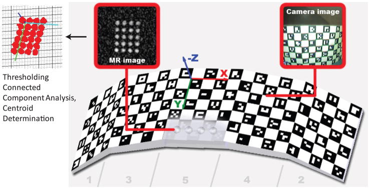
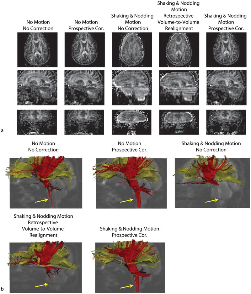
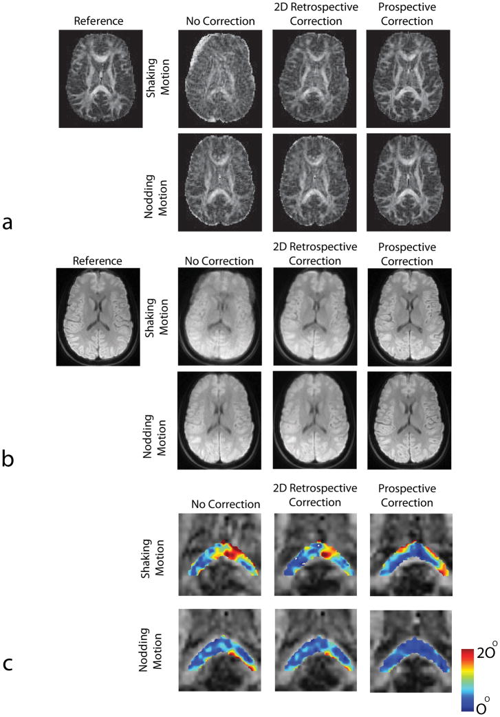
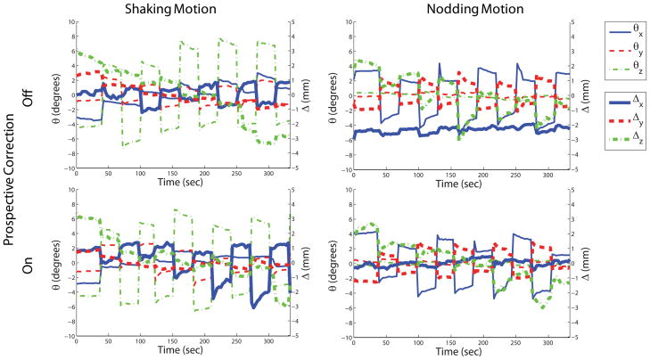
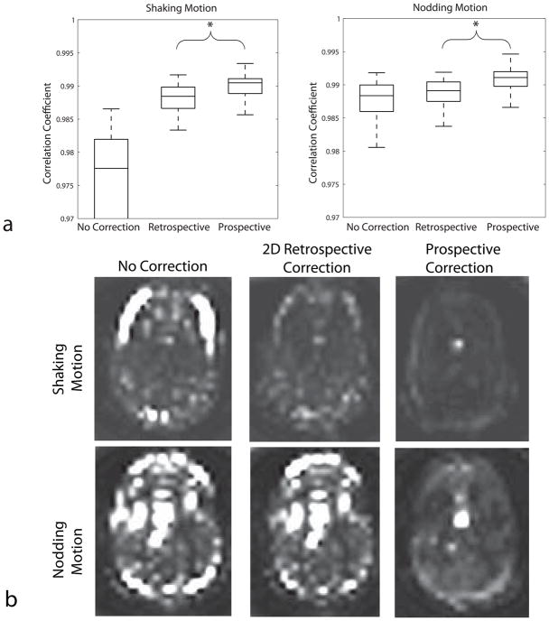
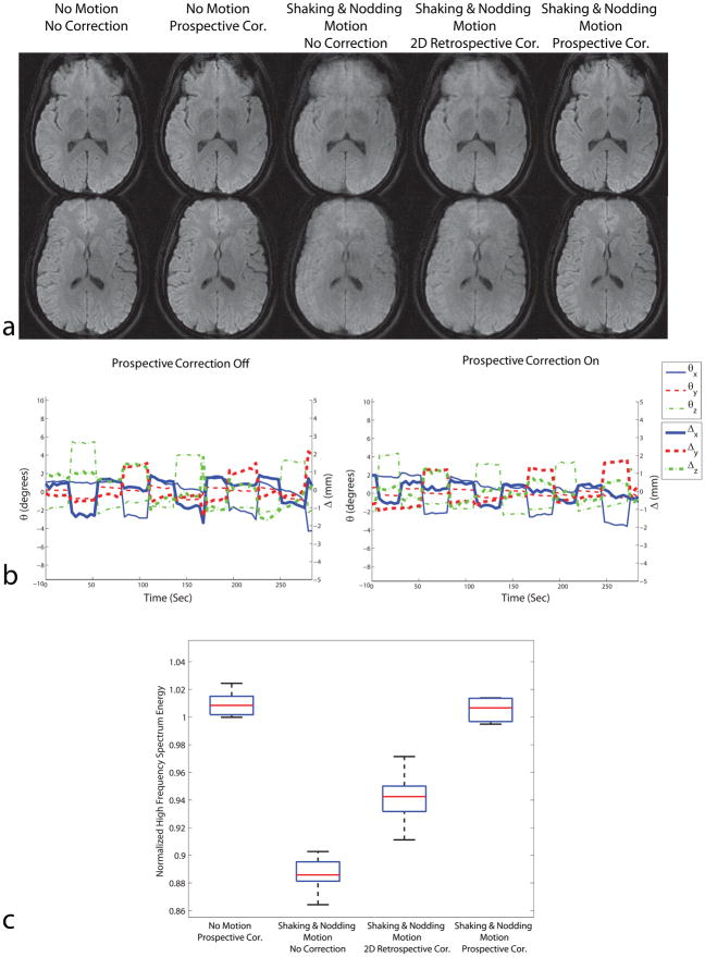
Head motion is a fundamental problem in brain MRI. The problem is further compounded in diffusion tensor imaging because of long acquisition times, and the sensitivity of the tensor computation to even small misregistration. To combat motion artifacts in diffusion tensor imaging, a novel real-time prospective motion correction method was introduced using an in-bore monovision system. The system consists of a camera mounted on the head coil and a self-encoded checkerboard marker that is attached to the patient's forehead. Our experiments showed that optical prospective motion correction is more effective at removing motion artifacts compared to image-based retrospective motion correction. Statistical analysis revealed a significant improvement in similarity between diffusion data acquired at different time points when prospective correction was used compared to retrospective correction (P<0.001).


Figures

Figure 1
Figure 1
System setup. An MR-compatible camera was mounted on the head coil inside the scanner bore (b). The camera took images of a self-encoded marker that was attached to the patient’s forehead (c,d). These images were processed by an external laptop where 1) the marker was segmented out, 2) its pose was estimated and 3) the 6 parameters (i.e. 3 rotations and 3 translations) to update the scanner geometry were sent to the scanner RF and gradient hardware controller. This allowed the slice being scanned to follow the subject’s head in real-time.
Figure 2
Figure 2
The 3D self-encoded marker used for pose tracking. Each black square on the marker included a unique 2D barcode that represented the position of that square on the marker geometry. In order to perform cross calibration, we also put MR-detectable agar droplets at the back of the marker. By measuring the poses of the MR detectable agar filled holes and the self-encoded checkerboard pattern simultaneously by the scanner and the camera, it was possible to find the relation between the scanner and the camera frame of references.
Figure 3
Figure 3
Results of in-vivo DTI experiments with 96×96 single-shot EPI readout. Reconstructed FA maps show that, even with retrospective volume-to-volume realignment, motion-related image artifacts remain because of intra-volume motion and spin history effects. Prospectively corrected FA maps show the most similarity with the reference dataset. The visualization of cortico-spinal tracts was also more successful when prospective correction was used compared to retrospectively corrected dataset.
Figure 4
Figure 4
Results of in-vivo DTI experiments with 128×128 resolution. Reconstructed FA maps (a) and isotropic DWI images (b) with and without 2D retrospective and prospective correction in the presence of shaking and nodding motion are shown. It can be seen that in the presence of random head motion the resulting images show significant motion related artifacts if optical motion tracking was turned off. These artifacts were largely removed after motion tracking was turned on. Superiority of prospective correction over retrospective method is better demonstrated in the presence of nodding motion. In this case, retrospective correction did not improve image quality because of the inability of this method to correct for through-plane motion, whereas prospectively corrected image looks very similar to the reference. The differences in structure between the reference FA map and the motion corrected maps are due to the fact that the subject did not return to the original position between “no motion” and “motion” scans. The angular deviation maps of the major eigenvectors from true orientations are shown in (c). True orientations are given by the dataset with no motion. Prospective correction provided eigenvectors with lower deviation compared to retrospective correction for both cases.
Figure 5
Figure 5
Motion plots corresponding to the shaking and nodding motion experiments in Fig. 4. The motion performed by the patient was similar for both scans when the motion tracking was turned off and on.
Figure 6
Figure 6
(a) Correlation coefficients of each navigator with the template for the three different methods. The higher correlation coefficient obtained while using prospective motion correction compared to retrospective correction implies that the navigator images are more “similar” to each other when prospective correction was used. (b) Pixel-by-pixel variance maps throughout the b=0 images (16 images). In the nodding motion case, prospective correction was much successful in reducing the variation between navigators. The bright spot visible in the lower 3 images is due to pulsation of the lateral ventricle.
Figure 7
Figure 7
Navigator signal loss due to motion. Due to the motion sensitivity of the diffusion weighting gradients, patient motion during the diffusion preparation period causes irrecoverable signal dropouts. This is clearly visible from the navigator signal that was obtained from the spiral-in readout. It can be seen that whenever there is patient motion (a), the navigator signal was significantly reduced. Only the dominant motion axes (i.e., θx and Δx) are shown to simplify the graph. Example navigator images from one slice are shown at the right (c).
Figure 8
Figure 8
Results of high resolution DWI experiments with resolution 256×256. Similar to the in-vivo experiments with 128×128 resolution, prospective motion correction performed better compared to retrospective correction due to the existence of through-plane motion. The motion plots are shown in (b). The distribution of high frequency spectrum energy for 7 slices in the acquisition is shown in (c). For all slices shown, the images with prospective correction had higher energy at high frequencies compared to uncorrected or retrospectively corrected images, implying less motion artifacts.

References

    1. Rohde GK, Barnett AS, Basser PJ, Marenco S, Pierpaoli C. Comprehensive approach for correction of motion and distortion in diffusion-weighted MRI. Magn Reson Med. 2004;51(1):103–114. - PubMed
    1. Jiang S, Xue H, Counsell S, Anjari M, Allsop J, Rutherford M, Rueckert D, Hajnal JV. Diffusion tensor imaging (DTI) of the brain in moving subjects: application to in-utero fetal and ex-utero studies. Magn Reson Med. 2009;62(3):645–655. - PubMed
    1. Leemans A, Jones DK. The B-matrix must be rotated when correcting for subject motion in DTI data. Magn Reson Med. 2009;61(6):1336–1349. - PubMed
    1. Pipe JG. Motion correction with PROPELLER MRI: application to head motion and free-breathing cardiac imaging. Magn Reson Med. 1999;42(5):963–969. - PubMed
    1. Liu C, Bammer R, Kim DH, Moseley ME. Self-navigated interleaved spiral (SNAILS): application to high-resolution diffusion tensor imaging. Magn Reson Med. 2004;52(6):1388–1396. - PubMed

Publication types