. 2012 Jan;33(1):179-91.
doi: 10.1002/hbm.21203. Epub 2011 Mar 22.

White matter maturation in visual and motor areas predicts the latency of visual activation in children

Affiliations

White matter maturation in visual and motor areas predicts the latency of visual activation in children

Colleen Dockstader et al. Hum Brain Mapp. 2012 Jan.

Abstract

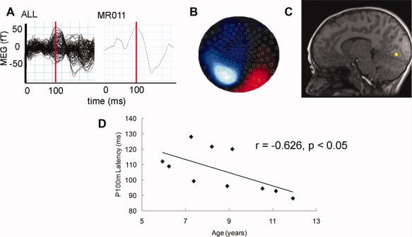
In humans, white matter maturation is important for the improvement of cognitive function and performance with age. Across studies the variables of white matter maturity and age are highly correlated; however, the unique contributions of white matter to information processing speed remain relatively unknown. We investigated the relations between the speed of the visually-evoked P100m response and the biophysical properties of white matter in 11 healthy children performing a simple, visually-cued finger movement. We found that: (1) the latency of the early, visually-evoked response was related to the integrity of white matter in both visual and motor association areas and (2) white matter maturation in these areas accounted for the variations in visual processing speed, independent of age. Our study is a novel investigation of spatial-temporal dynamics in the developing brain and provides evidence that white matter maturation accounts for age-related decreases in the speed of visual response. Developmental models of cortical specialization should incorporate the unique role of white matter maturation in mediating changes in performance during tasks involving visual processing.


Figures

Figure 1
Figure 1
Schematic depicting measured components in visual‐response task. Participants were instructed to attend to the cross on the screen and to abduct their right index finger as quickly as possible in response to the cross changing from white to green. Finger movements were confirmed with an EMG.
Figure 2
Figure 2
Images 2A through 2C depict data from a representative participant. (A) Evoked visual fields produced a well‐defined positive VEF around 100 ms following the visual cue. The evoked field was derived from the whole‐head MEG sensor data. (B) Preliminary localization on a contour map showed that distributions of magnetic fields appeared on either side of the occipital midline. (C) The dipole fit localized the early VEF response to the occipital region. Dipole fits for all participants are depicted in Supporting Information Figure 1. (D) There was a significant, negative correlation between age and the latency of the early VEF evoked visual response across participants.
Figure 3
Figure 3
(A) TBSS analyses determined regions where FA and early VEF latency were significantly related. Circled: Significant clusters within the right V5‐parietal region (first image) and right premotor region (second image) were isolated and masks were created to derive regional FA values for each participant which was then applied to a later hierarchical regression. (B) Correlational analyses for the overall FA Value and early VEF Latency revealed a significant inverse relationship between overall FA and early VEF latency where higher FA values predicted reduced latencies of the visual cortex.
Figure 4
Figure 4
V5/parietal SAM localizations. Spatial analysis of neural activations temporally‐related to the colour change of the visual stimulus. Nine of 11 subjects demonstrated visual stimulus‐related activations in the upper right parietal lobe, and 4 of these 9 also demonstrated stimulus‐related activations in the lower right parietal lobe. All of these activations are within the general region of the V5/parietal dorsal processing stream. [Color figure can be viewed in the online issue, which is available at wileyonlinelibrary.com.]
Figure 5
Figure 5
Premotor SAM localizations. Spatial analysis of neural activations temporally‐related to the colour change of the visual stimulus. Nine of 11 subjects demonstrated visual stimulus‐related activations in the right premotor region. [Color figure can be viewed in the online issue, which is available at wileyonlinelibrary.com.]
Figure 6
Figure 6
(A) Using TBSS we identified clusters of white matter in the parietal and frontal regions which FA significantly correlated with age. (B) There was a significant, positive correlation between age and the FA value extracted across all significant clusters. [Color figure can be viewed in the online issue, which is available at wileyonlinelibrary.com.]
Figure 7
Figure 7
(A) There was a significant inverse relationship between RT, as measured by EMG onset, and age whereby RT latency decreased with increasing age. (B) RT was positively correlated with the latency of the visual evoked response whereby RT decreases as the early VEF latency decreased.

Similar articles

Cited by

References

    1. Allison T, Wood CC, Goff WR ( 1983): Brain stem auditory, pattern‐reversal visual, and short‐latency somatosensory evoked potentials: Latencies in relation to age, sex, and brain and body size. Electroencephalogr Clin Neurophysiol 55: 619–636. - PubMed
    1. Als H, Duffy FH, McAnulty GB, Rivkin MJ, Vajapeyam S, Mulkern RV, Warfield SK, Huppi PS, Butler SC, Conneman N, Fischer C, Eichenwald EC ( 2004): Early experience alters brain function and structure. Pediatrics 113: 846–857. - PubMed
    1. Balachandran C, Klistorner AI, Billson F ( 2004): Multifocal VEP in children: Its maturation and clinical application. Br J Ophthalmol 88: 226–232. - PMC - PubMed
    1. Barnea‐Goraly N, Menon V, Eckert M, Tamm L, Bammer R, Karchemskiy A, Dant CC, Reiss AL ( 2005): White matter development during childhood and adolescence: A cross‐sectional diffusion tensor imaging study. Cereb Cortex 15: 1848–1854. - PubMed
    1. Barnikol UB, Amunts K, Dammers J, Mohlberg H, Fieseler T, Malikovic A, Zilles K, Niedeggen M, Tass PA ( 2006): Pattern reversal visual evoked responses of V1/V2 and V5/MT as revealed by MEG combined with probabilistic cytoarchitectonic maps. Neuroimage 31: 86–108. - PubMed

Publication types

LinkOut - more resources