. 2011 Nov;49(11):870-7.
doi: 10.1002/dvg.20750. Epub 2011 Aug 18.

Efficient inducible Cre-mediated recombination in Tcf21 cell lineages in the heart and kidney

Affiliations

Efficient inducible Cre-mediated recombination in Tcf21 cell lineages in the heart and kidney

Asha Acharya et al. Genesis. 2011 Nov.

Abstract

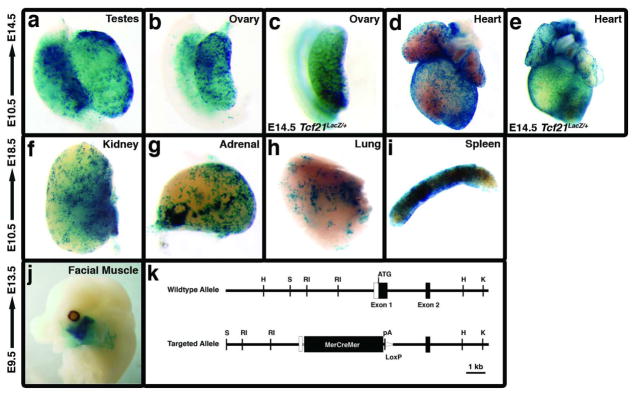
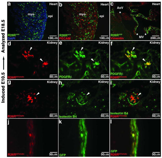
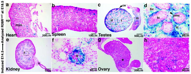
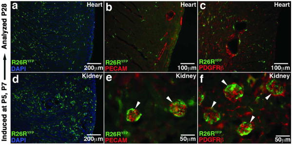
Tcf21 is a Class II bHLH family member with essential roles in the formation of the lungs, kidneys, gonads, spleen, and heart. Here, we report the utility of a mouse line with targeted insertion of a tamoxifen-inducible Cre recombinase, MerCreMer at the Tcf21 locus. This mouse line will permit the inducible expression of Cre recombinase in Tcf21-expressing cells. Using ROSA26 reporter mice, we show that Cre recombinase is specifically and robustly activated in multiple Tcf21-expressing tissues during embryonic and postnatal development. The expression profile in the kidney is particularly dynamic with the ability to cause recombination in mesangial cells at one time of induction and podocytes at another time. These features make the Tcf21-driven inducible Cre line (Tcf21(iCre) ) a valuable genetic tool for spatiotemporal gene function analysis and lineage tracing of cells in the heart, kidney, cranial muscle, and gonads.


Figures

Figure 1
Figure 1. Whole mount R26RLacZ tracing in Tcf21iCre induced embryonic tissues
(a,b and d) Whole mount views of β-galactosidase Cre reporter activity in E14.5 Tcf21iCre embryos tamoxifen-induced at E10.5. (a) male gonads (b) female gonads and (d) heart. (c and e) Comparative β-galactosidase expression in E14.5 Tcf21LacZ/+ ovary and heart respectively. (f–i) Cre reporter expression at E18.5 from embryos tamoxifen-induced at E10.5 (f) kidney (g) adrenal gland, (h) lung, and (i) spleen. (j) β-galactosidase activity in facial skeletal muscles of an E13.5 embryo tamoxifen-induced at E9.5. (k) Schematic representing wildtype and Tcf21iCre targeted locus. The MerCreMer fusion cDNA was knocked into the Tcf21 locus replacing the entire coding region of the first exon. Open boxes and grey line represent 5′ UTR and vector sequences respectively. H, HindIII; RI, EcoRI; S, SacI; K, KpnI; pA, polyadenylation signal sequence.
Figure 2
Figure 2. R26RYFP and R26RtdTomato tracing in Tcf21 iCre embryonic tissues induced at E10.5
(a–c) YFP reporter expression in E18.5 Tcf21iCre heart (green) and (d–i) R26RtdTomato tracing in E18.5 Tcf21iCre kidneys (red). (a) Epicardial and interstitial YFP reporter expression in embryonic heart costained with Dapi (blue) (b) Costain with PECAM (red) (c) Reporter expression in atrioventricular valves, costained with Vimentin (red). (d–i) Costainings (shown in green) with PDGFRβ(d–f) and endothelial marker Isolectin B4 (g–i) demonstrates labeling of mesangial cells of the glomeruli.. epi: epicardium; myo: myocardium; AoV: aortic valves; MV mitral valves marked by asterisk (*). Arrowheads in the kidney sections point to glomeruli. (j–l) Ex vivo migration assay showing Tcf21iCre labeled cells in the myocardium (red) are derived from AdGFP transduced (green) epicardial cells. Scale bars represent the indicated magnifications.
Figure 3
Figure 3. R26RLacZ tracing in E18.5 Tcf21 iCre embryonic tissues induced at E14.5
(a–h)β-galactosidase stained sections of E18.5 Tcf21iCre tissues tamoxifen- induced at E14.5. (a) heart, (b) spleen (c,d) testes, (e,f) kidney, and (g,h) ovary. (a) Reporter activity is seen in interstitial cells of the heart. (d) Magnified view of the testes showing reporter activity in peritubular myoid cells and interstitial cells of Leydig. β-galactosidase activity is seen in (f) glomerular podocytes. (h) magnified image of the ovary (region marked by asterisk (*) in (g)) showing β-galctosidase activity in thecal cells surrounding the follicles. Myo, myocardium; epi, epicardium; t, testes; m, mesonephrous; ce, ceolomic epithelium; tc, testis cords; pmc, peritubular myoid cells; ic, interstitial cells of leydig; bv, blood vessel; u, ureter; g, glomerulus. Scale bars represent the indicated magnifications.
Figure 4
Figure 4. R26RYFP tracing in Tcf21 iCre tissues following postnatal induction
(a–f) Cre activity detected by YFP reporter expression in postnatal day 28 (P28) Tcf21iCre tissues tamoxifen-induced at P5 and P7. (a–c) postnatal heart (d–f) kidney. YFP expression is detected by immunostaining (green). Costainings (shown in red) with PECAM (b and e) and PDGFRβ (c and f) highlight the blood vessels in the heart and glomerular capillaries and mesangial cells in the kidney respectively. Scale bars represent the indicated magnifications.

Similar articles

Cited by

References

    1. Bain G, Maandag ER, Izon DJ, Amsen D, Kruisbeek AM, Weintraub BC, Krop I, Schlissel MS, Feeney AJ, van Roon M, van der Valk M, te Riele HPJ, Berns A, Murre C. E2A proteins are required for proper B cell development and initiation of immunoglobin gene rearrangements. Cell. 1994;79:885–892. - PubMed
    1. Cui S, Ross A, Stallings N, Parker KL, Capel B, Quaggin SE. Disrupted gonadogenesis and male-to-female sex reversal in Pod1 knockout mice. Development. 2004;131:4095–4105. - PubMed
    1. Hidai H, Bardales R, Goodwin R, Quertermous T, Quertermous EE. Cloning of capsulin, a basic helix-loop-helix factor expressed in progenitor cells of the pericardium and the coronary arteries. Mech Dev. 1998;73:33–42. - PubMed
    1. Juhila J, Roozendaal R, Lassila M, Verbeek SJ, Holthofer H. Podocyte cell-specific expression of doxycycline inducible Cre recombinase in mice. J Am Soc Nephrol. 2005;17:648–654. - PubMed
    1. Lee JE, Hollenberg SM, Snider L, Turner DL, Lipnick L, Weintraub H. Conversion of Xenopus ectoderm into neurons by neuroD, a basic helix-loop-helix protein. Science. 1995;268:836–844. - PubMed

Publication types

MeSH terms

LinkOut - more resources