. 2011 Oct;66(4):1198-208.
doi: 10.1002/mrm.22906. Epub 2011 Mar 23.

32-channel RF coil optimized for brain and cervical spinal cord at 3 T

Affiliations

32-channel RF coil optimized for brain and cervical spinal cord at 3 T

J Cohen-Adad et al. Magn Reson Med. 2011 Oct.

Abstract

Diffusion and functional magnetic resonance imaging of the spinal cord remain challenging due to the small cross-sectional size of the cord and susceptibility-related distortions. Although partially addressable through parallel imaging, few highly parallel array coils have been implemented for the cervical cord. Here, we developed a 32-channel coil that fully covers the brain and c-spine and characterized its performance in comparison with a commercially available head/neck/spine array. Image and temporal signal-to-noise ratio were, respectively, increased by 2× and 1.8× in the cervical cord. Averaged g-factors at 4× acceleration were lowered by 22% in the brain and by 39% in the spinal cord, enabling 1-mm isotropic R = 4 multi-echo magnetization prepared gradient echo of the full brain and c-spine in 3:20 min. Diffusion imaging of the cord at 0.6 × 0.6 × 5 mm(3) resolution and tractography of the full brain and c-spine at 1.7-mm isotropic resolution were feasible without noticeable distortion. Improvements of this nature potentially enhance numerous basic and clinical research studies focused on spinal and supraspinal regions.


Figures

Figure 1
Figure 1
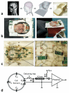
a. Methodology for optimal design of the coil using a 3D printer. Key steps were: automatic segmentation of an MPRAGE, reconstruction of 3D surface, design and printing of the coil frame. b. Photographs of the 30-channel coil with head and neck 2-channel paddles (although only one paddle can be combined at a time with conventional 32-channel MRI system). Switching between both paddles takes less than a minute. The coil fits all the 35 human subjects used for testing. c. Back of the coil array with zoomed panel focusing on the head/neck region. Preamplifiers were mounted close to each loop to minimize noise and cross-talk from cable currents. d. Schematics of a single array element, which consisted in a wire loop with four distributed capacitors. C4 was an adjusting capacitor for fine coil tuning.
Figure 2
Figure 2
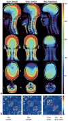
a. SNRcov-rSoS maps for the 32-channel coil with head paddle (left), the 32-channel with neck paddle (middle) and the standard coil (right) in one subject. For the two axial maps (two last rows), slices were oriented along the AC-PC for the brain and centered at C5 for the spine. SNR increase was visible in most regions of the brain and spinal cord with maximum gains within the cortex. SNR gain was quantitatively assessed in the brain, cerebellum, brainstem and cervical spinal cord. Results are showed in Table 2. b. Noise correlation coefficient matrices generated from acquisition without RF excitation. The averaged off-diagonal of the matrix was 7.7% for the 32-channel coil and 8.5% for the standard coil.
Figure 3
Figure 3
Left: Sagittal image showing the definition of ROIs used for quantifying the mean SNR in the supratentorial brain (1), cerebellum (2), brainstem (3) and cervical spinal cord (4). Middle, Right: Axial maps showing the ROIs used for quantifying the mean g-factor in the brain and spinal cord.
Figure 4
Figure 4
Maps of the inverse SENSE g-factor (1/g) at various accelerations for both coils. Closely fitted axial FOV was prescribed for the brain (top, FOV = 220 × 220 mm) and cervical region (bottom, FOV = 180 × 180 mm). For single acceleration direction (R=2 to 8), phase encoding was set to A-P. The peak g-factor is shown below each map. The spinal cord is circled in black. This figure suggests that 3x acceleration was feasible in the brain and spinal cord with relatively small cost in SNR. Note that there were not enough coil elements in the standard coil to accelerate with R=8 or R=3×3 in the cervical region.
Figure 5
Figure 5
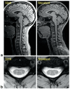
a. MEMPRAGE sequence. FOV = 320×224 mm, resolution = 1×1×1 mm3, TR/TI = 2200/1200ms, flip angle = 7°, R=4 (accelerated along A-P), BW = 285Hz/pix, TA = 3:20min. Images were not corrected for intensity non-uniformity and have the same color scaling. b. MEDIC sequence, transverse slice centered at C5 level. The same slice prescription was used for both coils. FOV = 154 mm, TR/TE = 1300/14 ms, flip angle = 30°, matrix = 256×256, resolution = 0.6×0.6×3 mm3, R=3, BW=260Hz/pix, saturation band covering the posterior neck, TA=2:31min.
Figure 6
Figure 6
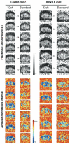
FA and maps of 95% angular confidence computed from FSL BedpostX for the 0.8×0.8 mm2 and 0.6×0.6 mm2 in-plane spatial resolution. Overall the 32-channel coil provided lower angular uncertainty in the whole cervical cord.
Figure 7
Figure 7
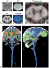
a. Example of DW images of the spinal cord at 0.6×0.6×5 mm3 spatial resolution. Dorsal/ventral roots were visible on the 2nd eigenvector. b. Full brain + c-spine tractography from 1.7mm isotropic DTI acquisition using the 32-channel coil with head paddle (74 sagittal slices, TR/TE = 14280/80 ms, R=3, b-value = 800 s/mm2, 30 diffusion-directions). No distortion correction was applied.
Figure 8
Figure 8
TSNR maps for the 32-channel (top) and standard (bottom) array coils for an axial slice centered at C4.

References

    1. Thurnher MM, Bammer R. Diffusion-weighted magnetic resonance imaging of the spine and spinal cord. Seminars in roentgenology. 2006;41(4):294–311. - PubMed
    1. Heidemann RM, Ozsarlak O, Parizel PM, Michiels J, Kiefer B, Jellus V, Müller M, Breuer F, Blaimer M, Griswold MA, Jakob PM. A brief review of parallel magnetic resonance imaging. Eur Radiol. 2003;13(10):2323–2337. - PubMed
    1. Kwong KK, Belliveau JW, Chesler DA, Goldberg IE, Weisskoff RM, Poncelet BP, Kennedy DN, Hoppel BE, Cohen MS, Turner R, Cheng H-M, Brady T, Rosen BR. Dynamic magnetic resonance imaging of human brain activity during primary sensory stimulation. Proc Natl Acad Sci U S A. 1992;89(12):5675–5679. - PMC - PubMed
    1. Stroman PW. Magnetic resonance imaging of neuronal function in the spinal cord: spinal FMRI. Clin Med Res. 2005;3(3):146–156. - PMC - PubMed
    1. Giove F, Garreffa G, Giulietti G, Mangia S, Colonnese C, Maraviglia B. Issues about the fMRI of the human spinal cord. Magn Reson Imaging. 2004;22(10):1505–1516. - PubMed

Publication types

MeSH terms