. 2011 Sep;164(1):170-80.
doi: 10.1111/j.1476-5381.2011.01336.x.

Thioredoxin interacting protein is a novel mediator of retinal inflammation and neurotoxicity

Affiliations

Thioredoxin interacting protein is a novel mediator of retinal inflammation and neurotoxicity

Mohammed M H Al-Gayyar et al. Br J Pharmacol. 2011 Sep.

Abstract

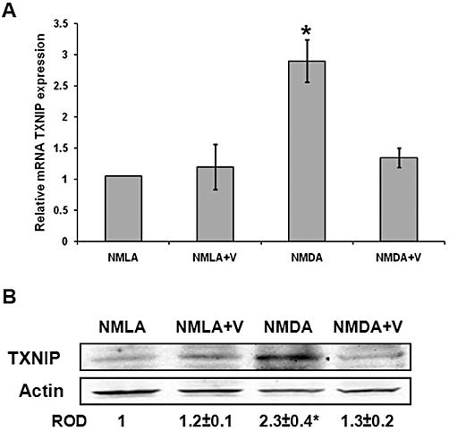
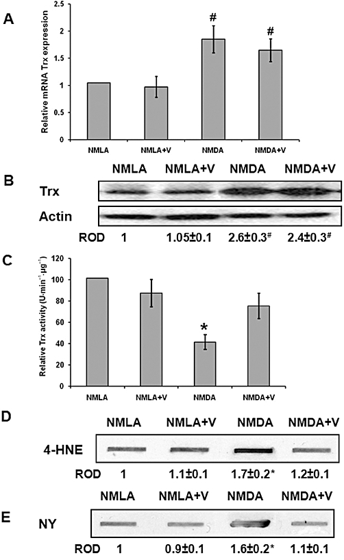
Background and purpose: Up-regulation of thioredoxin interacting protein (TXNIP), an endogenous inhibitor of thioredoxin (Trx), compromises cellular antioxidant and anti-apoptotic defences and stimulates pro-inflammatory cytokines expression, implying a role for TXNIP in apoptosis. Here we have examined the causal role of TXNIP expression in mediating retinal neurotoxicity and assessed the neuroprotective actions of verapamil, a calcium channel blocker and an inhibitor of TXNIP expression.

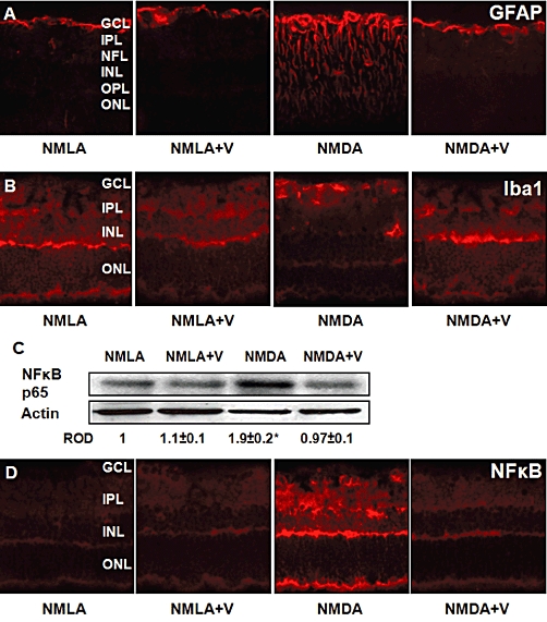
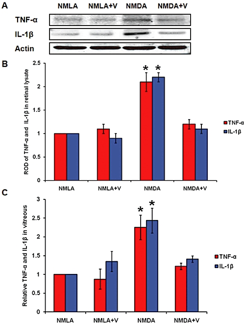
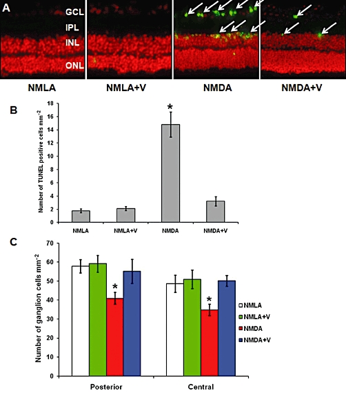
Experimental approach: Retinal neurotoxicity was induced by intravitreal injection of NMDA in Sprague-Dawley rats, which received verapamil (10 mg·kg(-1), p.o.) or vehicle. Neurotoxicity was examined by terminal dUTP nick-end labelling assay and ganglion cell count. Expression of TXNIP, apoptosis signal-regulating kinase 1 (ASK-1), NF-κB, p38 MAPK, JNK, cleaved poly-ADP-ribose polymerase (PARP), caspase-3, nitrotyrosine and 4-hydroxy-nonenal were examined by Western and slot-blot analysis. Release of TNF-α and IL-1β was examined by elisa.

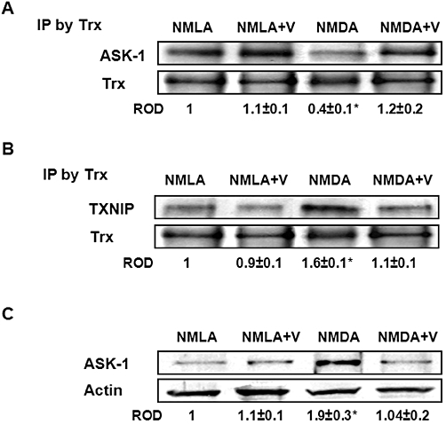
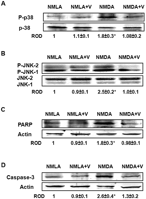
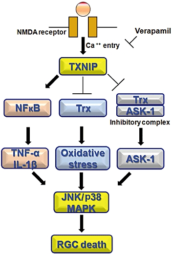
Key results: NMDA injection enhanced TXNIP expression, decreased Trx activity, causing increased oxidative stress, glial activation and release of TNF-α and IL-1β. Enhanced TXNIP expression disrupted Trx/ASK-1 inhibitory complex leading to release of ASK-1 and activation of the pro-apoptotic p38 MAPK/JNK pathway, as indicated by cleaved PARP and caspase-3 expression. Treatment with verapamil blocked these effects.

Conclusion and implications: Elevated TXNIP expression contributed to retinal neurotoxicity by three different mechanisms, inducing release of inflammatory mediators such as TNF-α and IL-1β, altering antioxidant status and disrupting the Trx-ASK-1 inhibitory complex leading to activation of the p38 MAPK/JNK apoptotic pathway. Targeting TXNIP expression is a potential therapeutic target for retinal neurodegenerative disease.


Figures

Figure 1
Figure 1
Verapamil blocks TXNIP expression in NMDA-injected retinas. (A) Real-time PCR analysis showed 2.9-fold increase in mRNA level of TXNIP in NMDA-injected rat retinas compared with NMLA-controls (n = 4–6). (B) Western blot analysis of rat retinal lysates showed 2.3-fold increase in TXNIP expression in NMDA-injected retinas, compared with NMLA-controls (n = 4–6). Treatment with verapamil (V; 10 mg·kg−1, p.o.) blocked TXNIP protein and mRNA level in NMDA injected retina but did not alter TXNIP in treated controls. Data expressed as fold value of control and represented as mean ± SEM. *P < 0.05, significantly different from the other groups. ROD, relative optical density.
Figure 2
Figure 2
Inhibiting TXNIP expression blocks activation of Müller and microglial cells and NF-κB expression. (A) Representative images showing a substantial increase in the intensity of GFAP immuno-reactivity in the filaments of Müller cells that extended from the nerve fiber layer (NFL) and inner plexiform layer (IPL) into the outer nuclear layer (ONL) of retina, compared with NMLA-controls (400 × magnification). (B) Representative images showing microglial activation as indicated by numerous Iba1-positive cells that appeared hypertrophic or amoeboid in the ganglion cell layer (GCL) and inner nuclear layer (INL) in rats injected with NMDA, compared with NMLA-controls (400 × magnification). (C) Western blot analysis of rat retinal lysate showing a 1.9-fold increase in NF-κB p65 in NMDA-injected retinas, compared with NMLA-controls (n = 6). Data expressed as fold value of control and represented as mean ± SEM. (D) Representative images showing prominent immunolocalization of NF-κB in the GCL, INL and outer plexiform layer (OPL), compared with NMLA-controls (400 × magnification). Treatment of rats with verapamil (V; 10 mg·kg−1, p.o.) blocked these effects in NMDA-injected rats but did not affect NMLA-controls. *P < 0.05, significantly different from the other groups.. ROD, relative optical density.
Figure 3
Figure 3
Inhibiting TXNIP expression blocks expression and release of inflammatory mediators. (A, B) Western blot analysis and statistical analysis of retinal lysate showed 2.1- and 2.2-fold increases in TNF-α and IL-Iβ, respectively, in NMDA-injected rats, compared with NMLA-controls (P < 0.05, n = 4–6). (C) Statistical analysis of the vitreous levels of TNF-α and IL-Iβ, using elisa. NMDA-injected retinas showed 2.2- and 2.4-fold increases in TNF-α and IL-Iβ, respectively, compared with NMLA-controls (n = 6–7). Treatment of rats with verapamil (V; 10 mg·kg−1, p.o.) blocked these effects in NMDA-injected rats but did not affect NMLA-controls. Data expressed as fold value of control and represented as mean ± SEM. *P < 0.05, significantly different from the other groups..; ROD, relative optical density.
Figure 4
Figure 4
Inhibiting TXNIP expression restores Trx activity and significantly reduces oxidative and nitrosative stress. (A) Real-time PCR analysis showed about two-fold increase in mRNA level of Trx in NMDA-injected rat retinas, compared with NMLA-controls, that was not reversed by verapamil (V) treatment (n = 4–6, data expressed as percent value of control and represented as mean ± SEM). (B) Western blot analysis showed 2.6-fold increase in the expression of Trx protein in NMDA-injected retinas, compared with NMLA-controls that was not reversed by verapamil treatment (n = 4). (C) Statistical analysis of Trx activity, NMDA injection caused 60% reduction in Trx activity as compared with NMLA-controls (n = 4–6, Data expressed as percent value of control and represented as mean ± SEM). (D, E) Slot-blot analysis of retinal lysate samples showed a 1.7-fold increase in 4-HNE adduct formation (a marker of oxidative stress) and a 1.6-fold increase in nitrotyrosine formation (a marker of peroxynitrite) in NMDA-injected rats compared with NMLA-controls (n = 4–6, data expressed as fold value of control and represented as mean ± SEM). Treatment with verapamil (V; 10 mg·kg−1, p.o.) blocked these effects in NMDA-injected rats but did not affect NMLA-controls. *P < 0.05, significantly different from the other groups.. #P < 0.05, significantly different from the controls. ROD, relative optical density.
Figure 5
Figure 5
Inhibiting TXNIP expression preserves the TRX-ASK-1 ‘inhibitory complex’ and prevents release of ASK-1. (A) Immunoprecipitation (IP) of Trx and immunoblotting with ASK-1 showed 60% reduction in interaction between Trx and ASK-1 in NMDA-injected rats, compared with NMLA-controls (n = 5). (B) Immunoprecipitation of Trx and immunoblotting with TXNIP showed a 1.6-fold increase in interaction between Trx and TXNIP in NMDA-injected retinas as compared with NMLA-control (n = 5). (C) Western blot analysis showing NMDA injection caused a 1.9-fold increase in ASK-1 expression in rat retina as compared with the NMLA-controls (n = 4–5). Co-treatment of animals with verapamil (V; 10 mg·kg−1, p.o.) blocked these effects in NMDA-injected rats but did not alter protein interaction in NMLA-controls. Data expressed as fold value of control and represented as mean ± SEM. *P < 0.05, significantly different from the other groups.. ROD, relative optical density.
Figure 6
Figure 6
Inhibiting TXNIP expression blocks activation of p38 MAPK/JNK apoptotic pathway. (A) Western blot analysis showed a 1.8-fold increase in p38 MAPK phosphorylation in NMDA-injected retinas as compared with NMLA-controls (n = 4–5). (B) Western blot analysis showing a 2.5-fold increase in phosphorylation of p-JNK-1 and p-JNK-2 in NMDA-injected retinas, compared with NMLA-controls (n = 4–6). (C, D) Western blot analysis showing 1.8- and 2.6-fold increases in cleaved PARP and cleaved caspase-3 expression, respectively, in NMDA-injected retinas, compared with NMLA-controls (n = 4). Co-treatment with verapamil (V; 10 mg·kg−1, p.o.) significantly reduced phosphorylation of p38 MAPK and p-JNK and blocked expression of cleaved PARP and cleaved caspase-3 in NMDA-injected animals but did not alter the basal levels in NMLA-controls. Data expressed as fold value of control and represented as mean ± SEM. *P < 0.05, significantly different from the other groups. ROD, relative optical density.
Figure 7
Figure 7
Inhibiting TXNIP expression prevents death and loss of RGC. (A, B) Representative images and statistical analysis showing that intravitreal injection of NMDA induced extensive RGC death as indicated by about 8-fold increase of TUNEL-labelled cells (arrows) mainly in RGC and inner nuclear layer (INL) of the rat retina compared with NMLA-controls (200 × magnification). (C) Statistical analysis of the number of neuronal cells in the ganglion cell layer (GCL) in retina sections stained with H/E. Intravitreal NMDA injections resulted in loss of 30% of cells in the GCL in the posterior and in the central regions of the retina. Co-treatment with verapamil (V) blocked these effects in NMDA-injected retinas but did not affect NMLA-injected controls. *P < 0.05, significantly different from the other groups..IPL, inner plexiform layer; ONL, outer nuclear layer; ROD, relative optical density.
Figure 8
Figure 8
Schematic representation of the mechanism of action of verapamil in neuroprotection. Enhanced TXNIP expression contributes to retinal neurotoxicity by multiple pathways: (A) inducing retinal inflammation as indicated by enhanced expression of NF-κB and release of pro-inflammatory cytokines, such as TNF-α and IL-1β; (B) modulating antioxidant defence and increasing oxidative stress; and (C) altering the inhibitory complex Trx/ASK-1 leading to activation of ASK-1. Together, these pathways can activate the apoptotic p38 MAPK/JNK pathway leading to RGC cell death.

Similar articles

Cited by

References

    1. Abdelsaid MA, Pillai BA, Matragoon S, Prakash R, Al-Shabrawey M, El-Remessy AB. Early intervention of tyrosine nitration prevents vaso-obliteration and neovascularization in ischemic retinopathy. J Pharmacol Exp Ther. 2010;332:125–134. - PubMed
    1. Al-Gayyar MMH, Abdelsaid MA, Matragoon S, Pillai BA, El-Remessy AB. Neuro and vascular protective effect of FeTPPs in NMDA-model: similarities to diabetes. Am J Pathol. 2010;177:1187–1197. - PMC - PubMed
    1. Ali TK, Matragoon S, Pillai BA, Liou GI, El-Remessy AB. Peroxynitrite mediates retinal neurodegeneration by inhibiting NGF survival signal in experimental and human diabetes. J Diabetes. 2008;57:889–898. - PubMed
    1. Ali TK, Al-Gayyar MMH, Matragoon S, Pillai BA, Abdelsaid MA, Nussbaum JJ, et al. Diabetes-induced peroxynitrite impairs the balance of ProNGF/NGF and causes neurovascular injury. Diabetologia. 2011;54:657–668. - PubMed
    1. Arkan MC, Hevener AL, Greten FR, Maeda S, Li ZW, Long JM, et al. IKK-beta links inflammation to obesity-induced insulin resistance. Nat Med. 2005;11:191–198. - PubMed

Publication types

MeSH terms