. 2011 Jun;218(6):660-7.
doi: 10.1111/j.1469-7580.2011.01366.x. Epub 2011 Mar 24.

The reinnervation and revascularisation pattern of scarless murine fetal wounds

Affiliations

The reinnervation and revascularisation pattern of scarless murine fetal wounds

James Henderson et al. J Anat. 2011 Jun.

Abstract

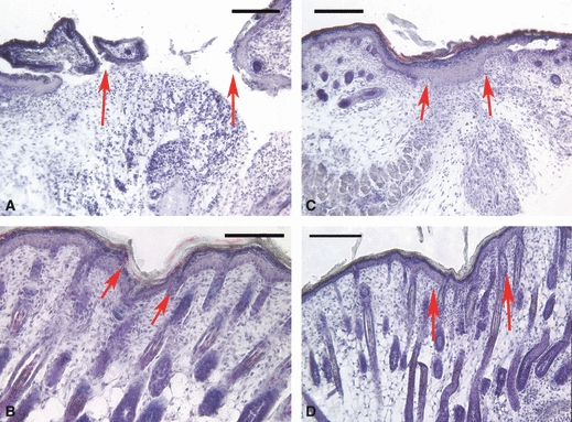
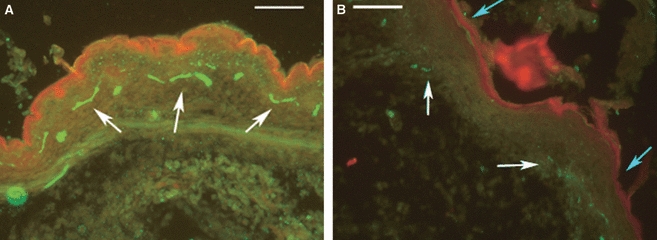
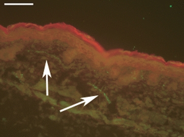
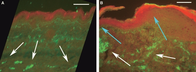
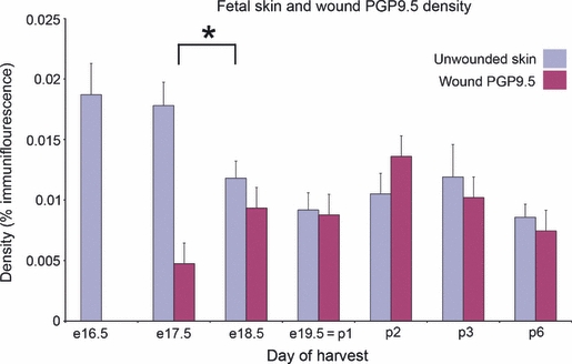
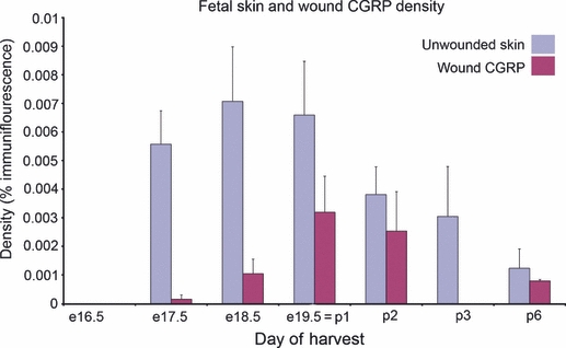
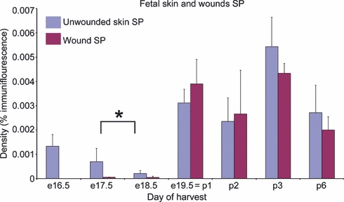
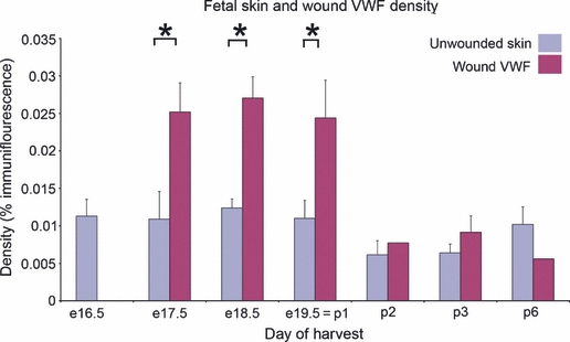
Fetal wounds can heal without scarring. There is evidence that the sensory nervous system plays a role in mediating inflammation and healing, and that the reinnervation pattern of adult wounds differs from that of unwounded skin. Ectoderm is required for development of the cutaneous nerve plexus in early gestation. It was hypothesised that scarless fetal wounds might completely regenerate their neural and vascular architecture. Wounds were made on mouse fetuses at embryonic day 16.5 of a 19.5-day gestation, which healed without visible scars. Immunohistochemical analysis of wound sites was performed to assess reinnervation, using antibodies to the pan neuronal marker PGP9.5 as well as to the neuropeptides calcitonin gene-related peptide (CGRP) and substance P (SP). Staining for the endothelial marker von Willebrand factor (VWF) allowed comparison of reinnervation and revascularisation. Wounds were harvested at timepoints from day 1 after wounding to postnatal day 6. Quantification of wound reinnervation and revascularisation was performed for timepoints up to 6 days post-wounding. Hypervascularisation of the wounds occurred within 24 h, and blood vessel density within the wounds remained significantly elevated until postnatal day 2 (4 days post- wounding), after which VWF immunoreactivity was similar between wound and control groups. Wound nerve density returned to a level similar to that of unwounded skin within 48 h of wounding, and PGP9.5 immunoreactive nerve fibre density remained similar to control skin thereafter. CGRP and SP immunoreactivity followed a similar pattern to that of PGP9.5, although wound levels did not return to those of control skin until postnatal day 1. Scarless fetal wounds appeared to regenerate their nerve and blood vessel microanatomy perfectly after a period of hypervascularisation.


Figures

Fig. 1
Fig. 1
Masson's stained section through a wound at embryonic days 17.5 (A), postnatal day 1 (B), postnatal day 3 (C), and postnatal day 6 (D). The scale bar is 100 μm in each, and the red arrows show the edges of the wound. By postnatal day 6, the wound has become almost impossible to distinguish from the surrounding skin. Although an increase in cellularity is apparent, it is difficult to be certain of the exact wound margins.
Fig. 2
Fig. 2
Immunostaining for the pan neuronal marker PGP9.5 in green. (A) Unwounded fetal skin from day e16.5. A cutaneous nervous plexus is present (white arrows). SP and CGRP immunoreactivity were not present at this stage of development. (B) A fetal wound at postnatal day 1, 3 days after wounding. PGP9.5 immunostaining shows nerve fibres in green (white arrow). In this wound they appear to be regenerating from the adjacent skin. The blue arrows indicate the wound edges. Scale bar: 100 μm.
Fig. 3
Fig. 3
Unwounded mouse skin from the day of birth (e19.5, which is the same as p1) showing immunostaining for CGRP (White arrows) that was only detected in very small amounts before this time. Scale bar: 100 μm.
Fig. 4
Fig. 4
Immunostaining for the vascular marker von Willebrand factor (VWF). (A) Unwounded fetal skin at day e16.5. A vascular plexus is seen (white arrows). (B) VWF immunostaining (green) in a fetal wound at postnatal day 1 (white arrows). The epithelium is counterstained red. The blue arrows show the wound margins, one of which is right at the edge of the field of view. Vessels appear to be regenerating from the wound edge at the right-hand side. Scale bar: 200 μm.
Fig. 5
Fig. 5
Histogram showing mean ± SEM reinnervation density of fetal wounds and control skin as indicated by PGP9.5 immunostaining at times after wounding. Wounds were made at day e16.5. A significant reduction in innervation density in control skin was seen between days e17.5 and e18.5 (*P < 0.05). Nerve density was significantly less in wounds 1 day after wounding (e17.5), but at all other timepoints, the density of nerve fibres was similar between wounds and control skin. Nerve density was measured as percentage immunofluorescence per high-power field after artefacts were excluded.
Fig. 6
Fig. 6
Histogram showing mean ± SEM reinnervation density of fetal wounds and control skin as indicated by CGRP immunostaining at times after wounding. Wounds were made at day e16.5, at which time no CGRP was detected in the fetal skin. A reduction in innervation density in control skin was seen between days p1 and p2. The CGRP innervation density of wounds was significantly lower than that of control skin for the first 2 days after wound creation; thereafter, CGRP density between control and wounded skin was similar. Nerve fibre density was measured as percentage immunofluorescence per high-power field after artefacts were excluded.
Fig. 7
Fig. 7
Histogram showing mean ± SEM reinnervation density of fetal wounds and control skin as indicated by SP immunostaining at times after wounding. Wounds were made at day e16.5. A significant reduction in innervation density in control skin was seen between day e17.5 and day e18.5 (*P < 0.05. Very little SP was detected in the fetal skin until the first postnatal day. SP levels were decreased in wounds on the first day. Otherwise, there was no difference in the density of SP in the wounds and control tissue at any timepoint.
Fig. 8
Fig. 8
Histogram showing mean ± SEM for fetal wound revascularisation and control skin as indicated by density of VWF immunoreactivity at times after wounding. Wounds were made at day e16.5. The hypervascularisation of wounds seen at days e17.5, e18.5 and p1 is statistically significant (*P < 0.05). VWF density was measured as percentage immunofluorescence per high-power field after artefacts were excluded.

Similar articles

Cited by

References

    1. Aldskogius H, Hermanson A, Jonsson CE. Reinnervation of experimental superficial wounds in rats. Plast Reconstr Surg. 1987;79:595–599. - PubMed
    1. Altun V, Hakvoort TE, van Zuijlen PP, et al. Nerve outgrowth and neuropeptide expression during the remodeling of human burn wound scars. A 7-month follow-up study of 22 patients. Burns. 2001;27:717–722. - PubMed
    1. Artuc M, Hermes B, Steckelings UM, et al. Mast cells and their mediators in cutaneous wound healing – active participants or innocent bystanders? Exp Dermatol. 1999;8:1–16. - PubMed
    1. Buckley G, Metcalfe AD, Ferguson MWJ. Peripheral nerve regeneration in the MRL/MpJ ear wound model. J Anat. 2011;218:163–172. doi: 10.1111/j.1469-7580.2010.01313.x. - DOI - PMC - PubMed
    1. Bush J, Duncan J, Bond J, et al. Scar-improving efficacy of Avotermin administered into the wound margins of skin incisions as evaluated by a randomised, double-blind, placebo-controlled, Phase II clinical trial. Plast Reconstr Surg. 2010;126:1604–1615. - PubMed

Publication types