Placentation in the eastern water skink (Eulamprus quoyii): a placentome-like structure in a lecithotrophic lizard
- PMID: 21434912
- PMCID: PMC3125902
- DOI: 10.1111/j.1469-7580.2011.01368.x
Placentation in the eastern water skink (Eulamprus quoyii): a placentome-like structure in a lecithotrophic lizard
Abstract
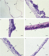
The eastern water skink (Eulamprus quoyii) has lecithotrophic embryos and was previously described as having a simple Type I chorioallantoic placenta. Indeed, it was the species upon which the definition of a Type I placenta was thought to be based, although we had cause to question that assumption. Hence we have described the morphology of the uterus of E. quoyii and found it to be more complex than previously supposed. The mesometrial pole of the uterus in E. quoyii displays a vessel-dense elliptical structure (the VDE) with columnar uterine epithelial cells. As pregnancy proceeds, the uterine epithelium near the mesometrial pole becomes folded and glands become hypertrophied, so that the morphology of VDE resembles that of a placentome, characteristic of Type III placentae. Unlike species with a Type III placenta, the apposing chorioallantoic membrane of E. quoyii is lined with squamous cells and interdigitates with the folded uterine epithelium. The remainder of the uterus is thin with a squamous uterine epithelium throughout pregnancy. Immunohistochemical localisation of blood vessels reveals a dense network of small capillaries directly beneath the folded epithelium of the VDE, while blood vessels are larger and sparser at the abembryonic pole of the uterus. Alkaline phosphatase (AP) activity is present in the uterine epithelium and sub-epithelial blood vessels in newly ovulated females. AP activity disappears from the epithelium between stages 27 and 29 of embryonic development and from the blood vessels after stage 34, but appears in the uterine glands at stage 35, where it remains until the end of pregnancy. Although the VDE is structurally similar to the placentomes found in other viviparous lizards, different distributions of AP activity in the uterus of E. quoyii and Pseudemoia spenceri suggest that the VDE may be functionally different from the placentome of the latter species. Our description of uterine morphology in E. quoyii provides evidence that, at least in some lineages, the evolution of a placentome may not occur in concert with the evolution of microlecithal eggs and obligate placentotrophy.
© 2011 The Authors. Journal of Anatomy © 2011 Anatomical Society of Great Britain and Ireland.
Figures


Similar articles
-
Morphology and development of the placentae in Eulamprus quoyii group skinks (Squamata: Scincidae).J Anat. 2012 May;220(5):454-71. doi: 10.1111/j.1469-7580.2012.01492.x. Epub 2012 Mar 16. J Anat. 2012. PMID: 22420511 Free PMC article.
-
Angiogenesis of the uterus and chorioallantois in the eastern water skink Eulamprus quoyii.J Exp Biol. 2010 Oct 1;213(Pt 19):3340-7. doi: 10.1242/jeb.046862. J Exp Biol. 2010. PMID: 20833927
-
Uterine epithelial changes during placentation in the viviparous skink Eulamprus tympanum.J Morphol. 2007 May;268(5):385-400. doi: 10.1002/jmor.10520. J Morphol. 2007. PMID: 17357138
-
A review of the evolution of viviparity in lizards: structure, function and physiology of the placenta.J Comp Physiol B. 2006 Mar;176(3):179-89. doi: 10.1007/s00360-005-0048-5. Epub 2005 Dec 7. J Comp Physiol B. 2006. PMID: 16333627 Review.
-
Structure, function, and evolution of the oviducts of squamate reptiles, with special reference to viviparity and placentation.J Exp Zool. 1998 Nov-Dec 1;282(4-5):560-617. J Exp Zool. 1998. PMID: 9867504 Review.
Cited by
-
Morphology and development of the placentae in Eulamprus quoyii group skinks (Squamata: Scincidae).J Anat. 2012 May;220(5):454-71. doi: 10.1111/j.1469-7580.2012.01492.x. Epub 2012 Mar 16. J Anat. 2012. PMID: 22420511 Free PMC article.
-
Anatomy of the female reproductive tract organs of the brown anole (Anolis sagrei).Anat Rec (Hoboken). 2024 Feb;307(2):395-413. doi: 10.1002/ar.25293. Epub 2023 Jul 28. Anat Rec (Hoboken). 2024. PMID: 37506227 Free PMC article.
-
Establishment and characterization of rough-tailed gecko original tail cells.Cytotechnology. 2018 Oct;70(5):1337-1347. doi: 10.1007/s10616-018-0223-7. Epub 2018 Jun 8. Cytotechnology. 2018. PMID: 29948549 Free PMC article.
-
The evolution of the placenta.Reproduction. 2016 Nov;152(5):R179-89. doi: 10.1530/REP-16-0325. Epub 2016 Aug 2. Reproduction. 2016. PMID: 27486265 Free PMC article. Review.
-
A review of the evolution of viviparity in squamate reptiles: the past, present and future role of molecular biology and genomics.J Comp Physiol B. 2011 Jul;181(5):575-94. doi: 10.1007/s00360-011-0584-0. Epub 2011 May 15. J Comp Physiol B. 2011. PMID: 21573966 Review.
References
-
- Adams SM, Biazik JM, Thompson MB, et al. Cyto-epitheliochorial placenta of the viviparous lizard Pseudemoia entrecasteauxii: a new placental morphotype. J Morphol. 2005;264:264–276. - PubMed
-
- Adams SM, Lui S, Jones SM, et al. Uterine epithelial changes during placentation in the viviparous skink Eulamprus tympanum. J Morphol. 2007;268:385–400. - PubMed
-
- Bansode FW, Chauhan SC, Makker A, et al. Uterine luminal epithelial alkaline phosphatase activity and pinopod development in relation to endometrial sensitivity in the rat. Contraception. 1998;58:61–68. - PubMed
-
- Biazik JM, Thompson MB, Murphy CR. The tight junctional protein occludin is found in the uterine epithelium of squamate reptiles. J Comp Physiol B. 2007;177:935–943. - PubMed
-
- Biazik JM, Thompson MB, Murphy CR. Claudin-5 is restricted to the tight junction region of uterine epithelial cells in the uterus of pregnant/gravid squamate reptiles. Anat Rec. 2008;291:547–556. - PubMed
Publication types
MeSH terms
Substances
LinkOut - more resources
Full Text Sources
Research Materials

