

The primary cilium of telocytes in the vasculature: electron microscope imaging
- PMID: 21435170
- PMCID: PMC4373428
- DOI: 10.1111/j.1582-4934.2011.01312.x
The primary cilium of telocytes in the vasculature: electron microscope imaging
Abstract
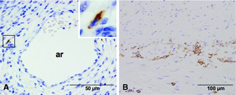
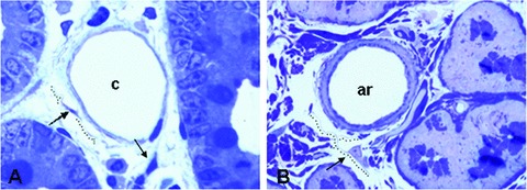
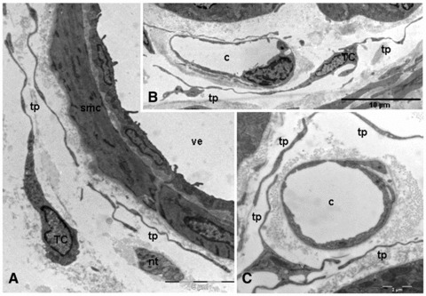
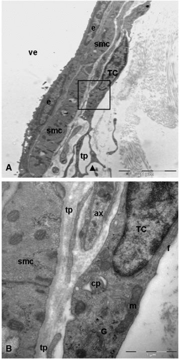
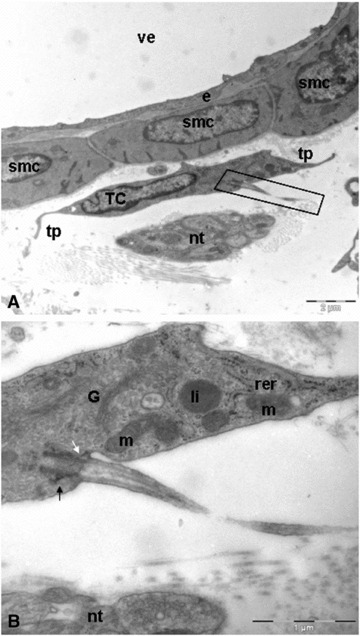
Blood vessels are highly organized and complex structure, which are far more than simple tubes conducting the blood to almost any tissue of the body. The fine structure of the wall of blood vessels has been studied previously using the electron microscope, but the presence the telocytes associated with vasculature, a specific new cellular entity, has not been studied in depth. Interestingly, telocytes have been recently found in the epicardium, myocardium, endocardium, human term placenta, duodenal lamina propria and pleura. We show the presence of telocytes located on the extracellular matrix of blood vessels (arterioles, venules and capillaries) by immunohistochemistry and transmission electron microscopy. Also, we demonstrated the first evidence of a primary cilium in telocytes. Several functions have been proposed for these cells. Here, the telocyte-blood vessels cell proximity, the relationship between telocytes, exosomes and nervous trunks may have a special significance.
© 2011 The Authors Journal of Cellular and Molecular Medicine © 2011 Foundation for Cellular and Molecular Medicine/Blackwell Publishing Ltd.
Figures


Similar articles
-
Telocytes in endocardium: electron microscope evidence.J Cell Mol Med. 2010 Sep;14(9):2330-4. doi: 10.1111/j.1582-4934.2010.01133.x. J Cell Mol Med. 2010. PMID: 20716125 Free PMC article.
-
Identification of telocytes in the lamina propria of rat duodenum: transmission electron microscopy.J Cell Mol Med. 2011 Jan;15(1):26-30. doi: 10.1111/j.1582-4934.2010.01207.x. J Cell Mol Med. 2011. PMID: 21054782 Free PMC article.
-
[Location of telocytes in mouse bronchial and pulmonary tissues].Zhonghua Bing Li Xue Za Zhi. 2012 Mar;41(3):172-5. doi: 10.3760/cma.j.issn.0529-5807.2012.03.006. Zhonghua Bing Li Xue Za Zhi. 2012. PMID: 22800480 Chinese.
-
Myocardial Telocytes: A New Player in Electric Circuitry of the Heart.Adv Exp Med Biol. 2016;913:241-251. doi: 10.1007/978-981-10-1061-3_16. Adv Exp Med Biol. 2016. PMID: 27796892 Review.
-
Vascular Telocytes.Adv Exp Med Biol. 2016;913:377-395. doi: 10.1007/978-981-10-1061-3_24. Adv Exp Med Biol. 2016. PMID: 27796900 Review.
Cited by
-
Role of Podoplanin-Positive Cells in Cardiac Fibrosis and Angiogenesis After Ischemia.Front Physiol. 2021 Apr 12;12:667278. doi: 10.3389/fphys.2021.667278. eCollection 2021. Front Physiol. 2021. PMID: 33912076 Free PMC article. Review.
-
Cellular Evidence of Telocytes as Novel Interstitial Cells Within the Magnum of Chicken Oviduct.Cell Transplant. 2017 Jan 24;26(1):135-143. doi: 10.3727/096368916X692942. Epub 2016 Sep 1. Cell Transplant. 2017. PMID: 27590447 Free PMC article.
-
Telocytes in human skin--are they involved in skin regeneration?J Cell Mol Med. 2012 Jul;16(7):1405-20. doi: 10.1111/j.1582-4934.2012.01580.x. J Cell Mol Med. 2012. PMID: 22500885 Free PMC article.
-
Tissue engineering and regenerative medicine -where do we stand?J Cell Mol Med. 2012 Jun;16(6):1157-65. doi: 10.1111/j.1582-4934.2012.01564.x. J Cell Mol Med. 2012. PMID: 22436120 Free PMC article. Review.
-
Telocytes in trachea and lungs.J Cell Mol Med. 2011 Oct;15(10):2262-8. doi: 10.1111/j.1582-4934.2011.01404.x. J Cell Mol Med. 2011. PMID: 21810171 Free PMC article.
References
-
- Eble JA, Niland S. The extracellular matrix of blood vessels. Curr Pharm Des. 2009;15:1385–400. - PubMed
-
- Skandalakis LJ, Skandalakis JE, Skandalakis PN. Surgical anatomy and technique: a pocket manual. 3. New York: Springer Science+Business Media LLC; 2009. Duodenum; pp. 333–46.
-
- Gabella G. Asymmetric distribution of dense bands in muscle cells of mammalian arterioles. J Ultrastruct Res. 1983;84:24–33. - PubMed
Publication types
MeSH terms
LinkOut - more resources
Full Text Sources
