Review
. 2011 Jul;50(3):267-77.
doi: 10.1016/j.plipres.2011.03.002. Epub 2011 Mar 22.

Role of membrane integrity on G protein-coupled receptors: Rhodopsin stability and function

Affiliations
Review

Role of membrane integrity on G protein-coupled receptors: Rhodopsin stability and function

Beata Jastrzebska et al. Prog Lipid Res. 2011 Jul.

Abstract

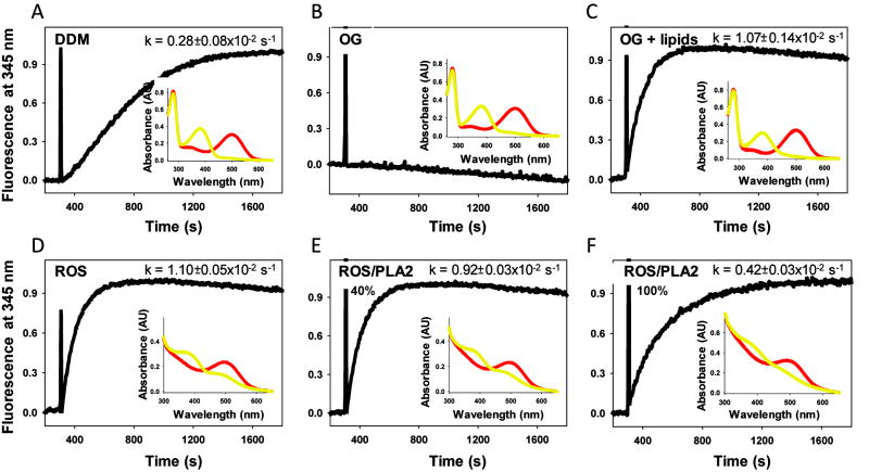
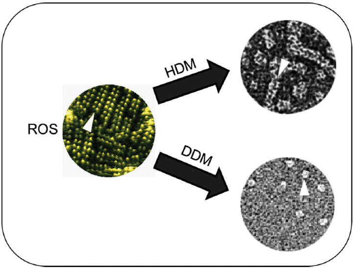
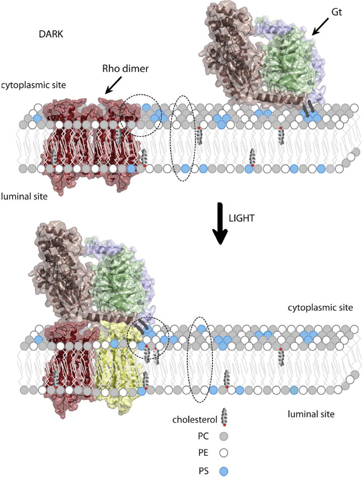
Rhodopsin is a prototypical G protein-coupled receptor (GPCR) - a member of the superfamily that shares a similar structural architecture consisting of seven-transmembrane helices and propagates various signals across biological membranes. Rhodopsin is embedded in the lipid bilayer of specialized disk membranes in the outer segments of retinal rod photoreceptor cells where it transmits a light-stimulated signal. Photoactivated rhodopsin then activates a visual signaling cascade through its cognate G protein, transducin or Gt, that results in a neuronal response in the brain. Interestingly, the lipid composition of ROS membranes not only differs from that of the photoreceptor plasma membrane but is critical for visual transduction. Specifically, lipids can modulate structural changes in rhodopsin that occur after photoactivation and influence binding of transducin. Thus, altering the lipid organization of ROS membranes can result in visual dysfunction and blindness.


Figures

Figure 1
Figure 1
Ability of different preparations of photoactivated rhodopsin to activate Gt (transducin). Increased intrinsic fluorescence of the Gtα subunit caused by interaction with photoactivated rhodopsin: (A) solubilized in dodecyl-β-D-maltoside (DDM); (B) solubilized in n-octyl-glucopyranoside (OG); (C) solubilized in OG and complemented with lipids isolated from ROS; (D) in ROS membranes; (E) in ROS membranes treated with phospholipase A2 (PLA2) at ~ 40 % digestion efficiency; (F) in ROS membranes treated with phospholipase A2 at ~ 100 % digestion efficiency. Rhodopsin (25 nM) was mixed with Gt (250 nM) and samples were illuminatedfor 30 s with a fiber light through band pass filter 480-520 nm. After 300 s of recording, 5 μM GTPγS was added. Reactions were carried out in a continuously stirred cuvette at 20°C in buffer composed of 20 mM BTP, 120 mM NaCl, 2 mM MgCl2 plus either 2 mM DDM or 30 mM OG. No detergent was present in the buffer when Gt was activated by ROS membranes. Relative activation rates were calculated from 3 independent experiments. Intrinsic fluorescence of Gtα was measured with a LS55 luminescence spectrophotometer (Perkin Elmer, Life Science), by using excitation and emission wavelengths of 300 nm and 345 nm, respectively (125, 126). Insets, Absorption spectra of rhodopsin solubilized in DDM (A), or in OG (B), or in OG and complemented with lipids isolated from ROS (C), rhodopsin in ROS membranes (D) and rhodopsin in ROS membranes treated with phospholipase A2 with ~ 40 % digestion efficiency (E), and rhodopsin in ROS membranes treated with phospholipase A2 with ~ 100 % digestion efficiency (F). Red lines represent spectra of ground state rhodopsin. Yellow lines represent spectra of photoactivated rhodopsin illuminated for 30 s.
Figure 2
Figure 2
Rhodopsin oligomerization. The propensity of different preparations of rhodopsin to oligomerize is illustrated. Atomic force microscopic (AFM) image of native disk membranes is colored green (left) and organization of rhodopsin molecules in dimers, packed in rows is shown by the arrowhead. Transmission electron microscopic (TEM) images of negatively stained ROS membranes solubilized in either hexyl-β-D-maltoside (HDM) or dodecyl-β-D-maltoside (DDM) are shown on the right. Arrowheads highlight whole rows of rhodopsin dimers extracted by HDM but just single rhodopsin dimers extracted by DDM.
Figure 3
Figure 3
Schematic organization of the retinal rod photoreceptor cell. A, Rod photoreceptor cell with labeled cell segments and major cellular elements. B, Membrane organization of a single disk showing location of rhodopsin dimers. C, Schematic representation of different states of rhodopsin after light activation.
Figure 4
Figure 4
Residues of rhodopsin interacting with specific lipids. (A) Aliphatic lipid chain of dipalmitoyl-sn-3-glycero-phosphatidylethanolamine (PE) interacting with cytoplasmic parts of H-VI and H-VII. (B) Cholesterol molecule interacting with two rhodopsin molecules in 2D crystals (antiparallel arrangement of rhodopsin molecules). Cholesterol interacts with the cytoplasmic side of H-IV and the extracellular sides of H-VI and H-VII. (C) Residues of rhodopsin preferentially interacting with DHA (a, a’), saturated fatty acids (b, b’), and cholesterol (c, c’). Primed panels are rotated 180°. The color scheme fo r helices of rhodopsin is: H-I - blue, H-II - cyan, H-III - green, H-IV - lime, H-V - yellow, H-VI - orange, H-VII - light red, H-8 - red. In A and B panels the lipids are shown with their hydrogen atoms; no lipids in panel C. In all panels the gray shadowing denotes rhodopsin residues interacting with lipids.
Figure 5
Figure 5
Reorganization of specific lipids in a disk membrane upon light illumination allows appropriate binding of Gt to photoexcited rhodopsin. Model of rhodopsin dimer (26, 39) is colored red in the dark state. Activated rhodopsin molecule is colored yellow. Gtα, Gtβ and Gtγ within heterotrimeric G protein are colored light pink, green and violet, respectively. Gt is attached to the membrane through myristoyl and farnesyl groups. Headgroups of phosphatidylcholine (PC), phosphatidylethanolamine (PE) and phosphatidylserine (PS) are depicted as small circles colored grey, white and blue, respectively. Movement of PS from the inner (lumenal) to the outer (cytoplasmic) leaflet of the lipid bilayer, as well as reorganization of PS, PE and cholesterol in close proximity to the activated rhodopsin molecule, is highlighted with a broken ellipsoid and circle, respectively. This lipid reorganization plays an important role in formation of the rhodopsin-Gt complex, thereby activating the visual signaling cascade.

Similar articles

Cited by

References

    1. Muller DJ, Wu N, Palczewski K. Vertebrate membrane proteins: structure, function, and insights from biophysical approaches. Pharmacol Rev. 2008;60:43–78. - PMC - PubMed
    1. Jacobson K, Mouritsen OG, Anderson RG. Lipid rafts: at a crossroad between cell biology and physics. Nat Cell Biol. 2007;9:7–14. - PubMed
    1. Palczewski K. G protein-coupled receptor rhodopsin. Annu Rev Biochem. 2006;75:743–767. - PMC - PubMed
    1. Smith SO. Structure and activation of the visual pigment rhodopsin. Annu Rev Biophys. 2010;39:309–328. - PubMed
    1. Aveldano MI. Phospholipid solubilization during detergent extraction of rhodopsin from photoreceptor disk membranes. Arch Biochem Biophys. 1995;324:331–343. - PubMed

Publication types

MeSH terms

LinkOut - more resources