Review
. 2011 Apr;178(4):1416-28.
doi: 10.1016/j.ajpath.2010.12.051.

Phagocytic clearance in neurodegeneration

Affiliations
Review

Phagocytic clearance in neurodegeneration

Jennifer D Sokolowski et al. Am J Pathol. 2011 Apr.

Abstract

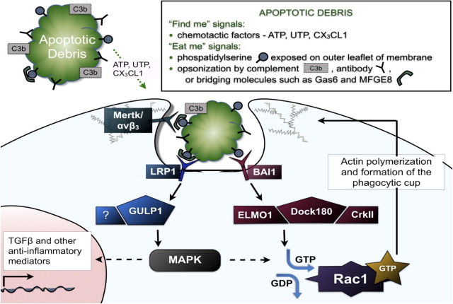
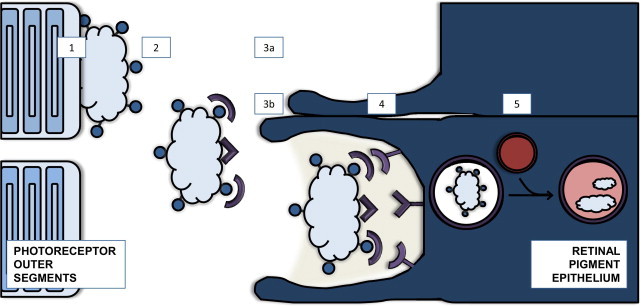
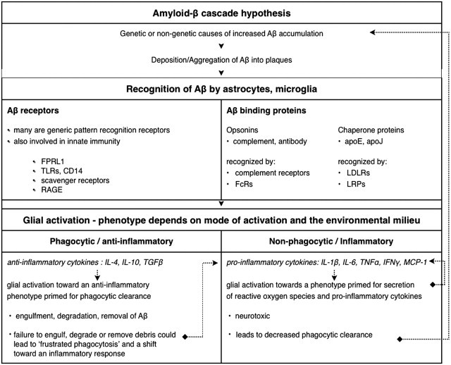
The cellular and molecular mechanisms of phagocytic clearance of apoptotic cells and debris have been intensely studied in invertebrate model organisms and in the mammalian immune system. This evolutionarily conserved process serves multiple purposes. Uncleared debris from dying cells or aggregated proteins can be toxic and may trigger exaggerated inflammatory responses. Even though apoptotic cell death and debris accumulation are key features of neurodegenerative diseases, relatively little attention has been paid to this important homeostatic function in the central nervous system (CNS). This review attempts to summarize our knowledge of phagocytic clearance in the CNS, with a focus on retinal degeneration, forms of which are caused by mutations in genes within known phagocytic pathways, and on Alzheimer's disease (AD). Interest in phagocytic clearance mechanisms in AD was stimulated by the discovery that immunization could promote phagocytic clearance of amyloid-β; however, much less is known about clearance of neuronal and synaptic corpses in AD and other neurodegenerative diseases. Because the regulation of phagocytic activity is intertwined with cytokine signaling, this review also addresses the relationships among CNS inflammation, glial responses, and phagocytic clearance.


Figures

Figure 1
Figure 1
Steps involved in phagocytosis of apoptotic debris. Apoptotic cells release diffusible find-me factors, such as ATP and chemokines, and express cell-surface eat-me signals, including phosphatidylserine. Complement and antibody molecules serve as opsonins for debris, and bridging molecules such as milk fat globule EGF factor 8 protein (MFGE8, also known as lactadherin) or growth arrest-specific protein 6 (GAS-6) bind to phosphatidylserine, targeting apoptotic debris for recognition. The phagocyte can thereby identify debris either by direct recognition of phosphatidylserine through receptors such as brain-specific angiogenesis inhibitor 1 (BAI1) or indirectly through recognition of opsonins/bridging molecules via lipoprotein receptor proteins (LRP), complement receptors, Fc receptors, αvβ3/5 integrin, and the receptor tyrosine kinase Mer (MerTK), among others. Signaling via these receptors is presumed to induce phagocytosis, but the precise players for all of these pathways have not yet been described. The small GTPase Rac1 has been implicated as a key downstream player responsible for regulating cytoskeletal alterations that are necessary for formation of the phagocytic cup and subsequent engulfment, and at least two signaling pathways upstream of Rac1 activation have been elucidated. First, a complex of ELMO-Dock180-CrkII acts downstream of the phosphatidylserine receptor, BAI1, and functions as a guanine exchange factor for Rac1. Second, LRP1 interacts with GULP, an adaptor protein that has been linked to Rac1 activation, possibly via mitogen-activated protein kinase (MAPK). Depending on the context, signaling may lead not only to phagocytosis, but also to changes in cell morphology, induction of migration, and secretion of cytokines. For example, engulfment of apoptotic debris stimulates production of anti-inflammatory mediators, such as transforming growth factor β (TGF-β).
Figure 2
Figure 2
Sequence of events and factors involved in clearance of photoreceptor outer segments (POS) in the retina. Photoreceptors shed their tips diurnally in response to light, and the retinal pigment epithelium (RPE) is responsible for clearing this cellular debris. Retinal degeneration can occur as a result of defects in modification, recognition, engulfment, or degradation of photoreceptor debris. 1: Membrane modification. ABCA4 functions in removal of unmetabolizable byproducts such as all-trans retinal and N-retinylidene-PE on the POS membranes. 2: Segments are shed and expose phosphatidylserine. 3: Opsonization. 3a: GAS-6, protein S, and MFGE8 opsonize the shed POS by binding phosphatidylserine. 3b: Tubby and tubby-like protein 1 are ligands for receptor tyrosine kinase MerTK and promote phagocytosis via an unknown target, independent of PS. 4: Receptor recognition and engulfment. Receptors such as MerTK, lipid scavenger receptor CD36, integrin adhesion receptor (αvβ5), and TYRO3 recognize the shed POS and promote engulfment. 5: Intracellular trafficking of phagosomes. Phagosome-lysosome fusion leads to degradation of engulfed material. Mutations in genes involved in these pathways that have been linked to human disease include ABCA4 (Stargardt's macular degeneration), MERTK (retinitis pigmentosa), and MYO7A (Usher's syndrome).
Figure 3
Figure 3
Sequence of events connecting the amyloid cascade hypothesis to putative glial cell responses. Receptors that stimulate phagocytosis are also involved in inducing inflammatory pathways, and clearly the mechanism of activation determines the activation phenotype of the cell. It has been posited that frustrated phagocytosis may occur when glia are unable to clear debris such as Aβ. Persistence of extracellular debris leads to continual stimulation and production of degrading enzymes and reactive oxygen species, as well as proinflammatory signals. This chronic inflammation creates an environment that is toxic to neurons and promotes neurodegeneration. In theory, tipping the balance to improve phagocytic clearance and decrease secretion of proinflammatory mediators may ameliorate Aβ pathology. The dotted lines illustrate the concept that pathology may be amplified due to a pro-inflammatory milieu and/or defects in phagocytic clearance.

References

    1. Elliott M.R., Ravichandran K.S. Clearance of apoptotic cells: implications in health and disease. J Cell Biol. 2010;189:1059–1070. - PMC - PubMed
    1. Lee C.Y., Landreth G.E. The role of microglia in amyloid clearance from the AD brain. J Neural Transm. 2010;117:949–960. - PMC - PubMed
    1. Cahoy J.D., Emery B., Kaushal A., Foo L.C., Zamanian J.L., Christopherson K.S., Xing Y., Lubischer J.L., Krieg P.A., Krupenko S.A., Thompson W.J., Barres B.A. A transcriptome database for astrocytes, neurons, and oligodendrocytes: a new resource for understanding brain development and function. J Neurosci. 2008;28:264–278. - PMC - PubMed
    1. DeWitt D.A., Perry G., Cohen M., Doller C., Silver J. Astrocytes regulate microglial phagocytosis of senile plaque cores of Alzheimer's disease. Exp Neurol. 1998;149:329–340. - PubMed
    1. Takuma K., Fang F., Zhang W., Yan S., Fukuzaki E., Du H., Sosunov A., McKhann G., Funatsu Y., Nakamichi N., Nagai T., Mizoguchi H., Ibi D., Hori O., Ogawa S., Stern D.M., Yamada K., Yan S.S. RAGE-mediated signaling contributes to intraneuronal transport of amyloid-beta and neuronal dysfunction. Proc Natl Acad Sci USA. 2009;106:20021–20026. - PMC - PubMed

Publication types