. 2011 Apr;178(4):1461-9.
doi: 10.1016/j.ajpath.2010.12.026.

Prognostic significance of TRIM24/TIF-1α gene expression in breast cancer

Affiliations

Prognostic significance of TRIM24/TIF-1α gene expression in breast cancer

Monique Chambon et al. Am J Pathol. 2011 Apr.

Abstract

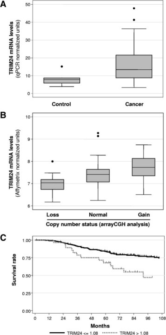
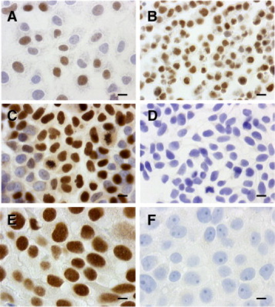
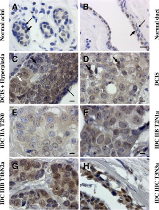
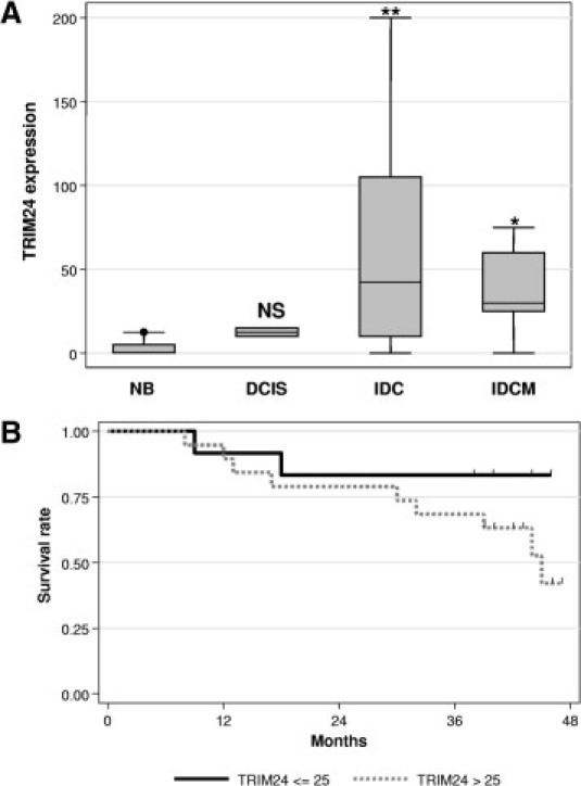
In this study, we have analyzed the expression of TRIM24/TIF-1α, a negative regulator of various transcription factors (including nuclear receptors and p53) at the genomic, mRNA, and protein levels in human breast tumors. In breast cancer biopsy specimens, TRIM24/TIF-1α mRNA levels (assessed by Real-Time Quantitative PCR or microarray expression profiling) were increased as compared to normal breast tissues. At the genomic level, array comparative genomic hybridization analysis showed that the TRIM24/TIF-1α locus (7q34) exhibited both gains and losses that correlated with mRNA levels. By re-analyzing a series of 238 tumors, high levels of TRIM24/TIF-1α mRNA significantly correlated with various markers of poor prognosis (such as the molecular subtype) and were associated with worse overall survival. By using a rabbit polyclonal antibody for immunochemistry, the TRIM24/TIF-1α protein was detected in nuclei of normal luminal epithelial breast cells, but not in myoepithelial cells. Tissue microarray analysis confirmed that its expression was increased in epithelial cells from normal to breast infiltrating duct carcinoma and correlated with worse overall survival. Altogether, this work is the first study that shows that overexpression of the TRIM24/TIF-1α gene in breast cancer is associated with poor prognosis and worse survival, and it suggests that this transcription coregulator may play a role in mammary carcinogenesis and represent a novel prognostic marker.


Figures

Figure 1
Figure 1
Expression of TRIM24/TIF-1α mRNA in breast carcinomas. A: TRIM24/TIF-1α mRNA levels were determined on 11 normal breast tissues (Control) and 135 breast carcinomas by RT-QPCR as described in Materials and Methods. Data were normalized using the IPO8 and HBMS mRNAs and are presented as box-plots showing the median value, upper and lower quartiles together and minimum and maximum non-atypical data values. B: TRIM24/TIF-1α mRNA levels were determined by Affymetrix microarray analysis (Affymetrix probe set 213301_x_at) in a series of 108 breast tumors with losses (13 cases), gains (18 cases), or no alterations (77 cases) at the corresponding locus as determined by aCGH analysis. Data are represented as box-plots. C: Kaplan–Meier analysis of overall survival according to TRIM24/TIF-1α mRNA expression in the 238-tumor set from Fan et al. Patients with high TRIM24/TIF-1α mRNA expression (expression level more than the optimal threshold of 1.08 - gray solid line) were compared to patients with low TRIM24/TIF-1α mRNA expression (expression level less than or equal to the optimal threshold of 1.08 - black solid line). Differences in survival rates were statistically significant when compared using a log-rank test (P < 0.001). Tick marks indicate censored events.
Figure 2
Figure 2
TRIM24/TIF-1α protein detection in breast cancer cells and tissues. Immunohistochemistry staining of the TRIM24/TIF-1α protein was performed as described in Materials and Methods after incubation with TIF-1α (TS1) polyclonal primary antibody (1/3000) at room temperature. A: MCF7 cells grown on glass coverslips. B: Paraffin-embedded MCF7 cell pellet sections. C, D: MCF7 cells grown on glass coverslips were incubated with TS1 antibody after incubation (D) or not (C) with 15 μg/mL of the GST-TRIM24/TIF-1α fusion protein used for immunization. E: Paraffin-embedded human breast IDC cells. F: Negative controls were performed with the rabbit pre-immune serum. (Original magnification X 630; scale bars = 10 μm.)
Figure 3
Figure 3
TRIM24/TIF-1α expression in human normal and tumoral tissues. Normal and tumor breast samples of the CBA2 tissue array were immunostained with the rabbit polyclonal TS1 antibody. Normal acini (A) and a normal duct (B) showing weak staining in some nuclei of epithelial cells (arrows). C–H: Representative lesions from six cases of the CBA2 tissue array showing increased intensity of staining with the TS1 antibody. C: Weak staining in a hyperplasic area (shown by a black short arrow) beside unlabeled myoepithelial cells (long black arrow) and labeled intraductal cancerous cells (white short arrow). D: Ductal carcinoma in situ (DCIS) where only two nuclei appear weakly labeled (arrows). E–H: Increase in nuclear staining observed in IDC with different staging (E) stage IIA, N0; (F) stage IIB, N1; (G) stage IIIB, N2; and (H) stage IIIC, N3. Case numbers correspond to an individual patient. (Original magnification X 630; scale bars = 10 μm.)
Figure 4
Figure 4
Tissue array analysis of TRIM24/TIF-1α expression. A: Data obtained after quantification of the TRIM24/TIF-1α protein by immunochemistry using the TS1 antibody on the CBA2 tissue array were represented as box-plots. The labeling values were plotted according to the different groups: normal breast (NB), DCIS, IDC, and IDCM. Statistical analysis was performed using the Mann-Whitney test with reference to the NB group data (*P < 0.05, **P < 0.01), NS. B: Overall survival was estimated for the 31 patients analyzed in the CBA2 tissue array using the Kaplan-Meier method according to TRIM24/TIF-1α protein expression. Patients with high TRIM24/TIF-1α expression (a level greater than the optimal threshold of 25 - gray solid line) were compared to patients with low TRIM24/TIF-1α expression (less than or equal to the optimal threshold of 25 - black solid line). The log-rank method was used to test the difference between the two groups, and patients with high levels of TRIM24/TIF-1α expression had significantly worse overall survival (P = 0.025). Tick marks indicate censored events.

References

    1. Le Douarin B., Zechel C., Garnier J.M., Lutz Y., Tora L., Pierrat P., Heery D., Gronemeyer H., Chambon P., Losson R. The N-terminal part of TIF1, a putative mediator of the ligand-dependent activation function (AF-2) of nuclear receptors, is fused to B-raf in the oncogenic protein T18. EMBO J. 1995;14:2020–2033. - PMC - PubMed
    1. Thenot S., Henriquet C., Rochefort H., Cavailles V. Differential interaction of nuclear receptors with the putative human transcriptional coactivator hTIF1. J Biol Chem. 1997;272:12062–12068. - PubMed
    1. Le Douarin B., Nielsen A.L., Garnier J.M., Ichinose H., Jeanmougin F., Losson R., Chambon P. A possible involvement of TIF1 alpha and TIF1 beta in the epigenetic control of transcription by nuclear receptors. EMBO J. 1996;15:6701–6715. - PMC - PubMed
    1. Le Douarin B., You J., Nielsen A.L., Chambon P., Losson R. TIF1alpha: a possible link between KRAB zinc finger proteins and nuclear receptors. J Steroid Biochem Mol Biol. 1998;65:43–50. - PubMed
    1. Teyssier C., Ou C.Y., Khetchoumian K., Losson R., Stallcup M.R. Transcriptional intermediary factor 1 alpha mediates physical interaction and functional synergy between the coactivator-associated arginine methyltransferase 1 and glucocorticoid receptor-interacting protein 1 nuclear receptor coactivators. Mol Endocrinol. 2006;20:1276–1286. - PMC - PubMed

MeSH terms