. 2011 Mar 24;69(6):1061-8.
doi: 10.1016/j.neuron.2011.02.040.

Effects and mechanisms of wakefulness on local cortical networks

Affiliations

Effects and mechanisms of wakefulness on local cortical networks

Christine M Constantinople et al. Neuron. .

Abstract

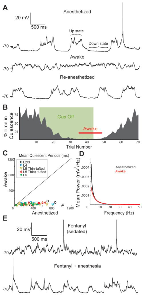
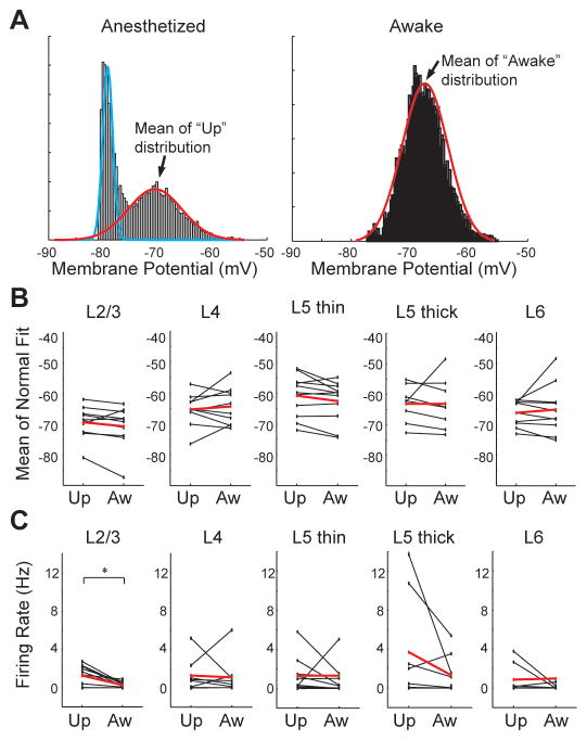
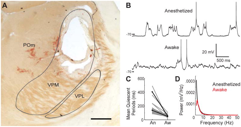
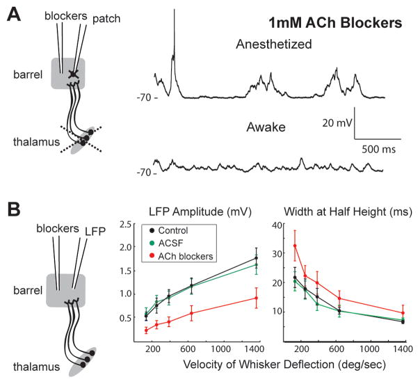
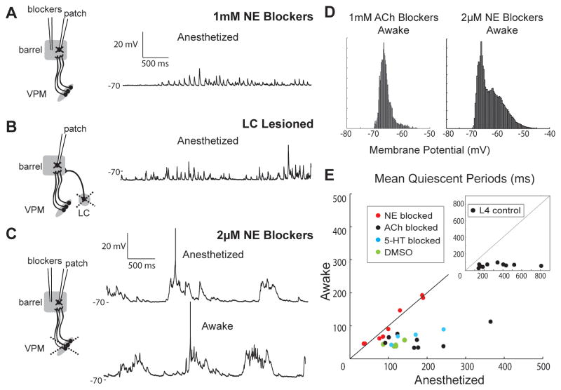
Mammalian brains generate internal activity independent of environmental stimuli. Internally generated states may bring about distinct cortical processing modes. To investigate how brain state impacts cortical circuitry, we recorded intracellularly from the same neurons, under anesthesia and subsequent wakefulness, in rat barrel cortex. In every cell examined throughout layers 2-6, wakefulness produced a temporal pattern of synaptic inputs differing markedly from those under anesthesia. Recurring periods of synaptic quiescence, prominent under anesthesia, were abolished by wakefulness, which produced instead a persistently depolarized state. This switch in dynamics was unaffected by elimination of afferent synaptic input from thalamus, suggesting that arousal alters cortical dynamics by neuromodulators acting directly on cortex. Indeed, blockade of noradrenergic, but not cholinergic, pathways induced synaptic quiescence during wakefulness. We conclude that global brain states can switch local recurrent networks into different regimes via direct neuromodulation.


Figures

Figure 1
Figure 1. Wakefulness abolishes periods of synaptic quiescence in all cortical layers
(A) In vivo whole-cell recording of a L2/3 pyramidal neuron while a rat was anesthetized (top), awake (middle) and re-anesthetized (bottom). (B) Percentage of each 5-second trial that the cell spent during periods of synaptic quiescence. (C) Scatterplot of the mean lengths of quiescent periods during wakefulness and anesthesia. (D) Average Vm power spectra for all neurons during anesthesia and wakefulness. (E) A L4 neuron under fentanyl sedation and subsequent administration of anesthetic.
Figure 2
Figure 2. The awake state is a persistent up-like state
(A) Vm histograms of a L5 pyramidal neuron during anesthesia and wakefulness, fit by a mixture of normals and a single normal, respectively. (B) Summary plots comparing the mean of the up and awake distributions for 52 cells (black, individual cells; red, means). (C) Summary plots comparing the firing rates during up states (under anesthesia) and awake periods.
Figure 3
Figure 3. Thalamus is not required for producing an awake Vm
(A) Coronal section showing an electrolytic VPM lesion, centered on the C2 barreloid. Scale bar, 500 μm. (B) A L4 star pyramid in the C2 barrel following the lesion shown in A. (C) Mean length of quiescent periods in L4 neurons following lesions. (D) Average Vm power spectra of lesion-only data in C.
Figure 4
Figure 4. ACh is not required for awake Vm but modifies sensory responses
(A) Left, schematic of experiment combining thalamic lesion with local perfusion of 1 mM blockers of muscarinic and nicotinic ACh receptors. Right, ACh blockers do not prevent transition to awake Vm patterns in example L4 star pyramid. (B) Local perfusion of blockers during LFP recording with thalamus intact (left). ACh blockers but not aCSF vehicle suppress amplitudes (middle) and extend durations (width at half amplitude) of whisker-evoked LFPs across velocities (right).
Figure 5
Figure 5. Blocking NE prevents awake Vm and induces synaptic quiescence
(A) L4 star pyramid in the presence of 1 mM NE blockers (thalamus intact). (B) L4 star pyramid after lesion of ipsilateral locus coeruleus (thalamus intact, no blockers). (C) L4 spiny stellate during local perfusion of 2 μM NE blockers after thalamic lesion. (D) Vm histograms of the cells shown in Figures 4A (left) and 5C (right) during wakefulness. (E) Scatter plot of the mean quiescent periods of L4 neurons during wakefulness and anesthesia in the presence of NE, ACh or 5-HT1A/B blockers or DMSO vehicle following thalamic lesion. Inset, L4 control data (from Figure 1C).

References

    1. Aston-Jones G, Bloom FE. Activity of norepinephrine-containing locus coeruleus neurons in behaving rats anticipates fluctuations in the sleep-waking cycle. J Neurosci. 1981;1:876–886. - PMC - PubMed
    1. Berridge CW, Page ME, Valentino RJ, Foote SL. Effects of locus coeruleus inactivation on electroencephalographic activity in neocortex and hippocampus. Neuroscience. 1993;55:381–393. - PubMed
    1. Blake H, Gerard RW. Brain potential during sleep. American Journal of Physiology. 1937;119:692–702.
    1. Blanco-Centurion C, Gerashchenko D, Salin-Pascual RJ, Shiromani PJ. Effects of hypocretin2-saporin and antidopamine-beta-hydroxylase-saporin neurotoxic lesions of the dorsolateral pons on sleep and muscle tone. Eur J Neurosci. 2004;19:2741–2752. - PMC - PubMed
    1. Briand LA, Gritton H, Howe WM, Young DA, Sarter M. Modulators in concert for cognition: modulator interactions in the prefrontal cortex. Prog Neurobiol. 2007;83:69–91. - PMC - PubMed

Publication types