. 2011 Mar 25;34(3):303-14.
doi: 10.1016/j.immuni.2011.03.004.

RUNX transcription factor-mediated association of Cd4 and Cd8 enables coordinate gene regulation

Affiliations

RUNX transcription factor-mediated association of Cd4 and Cd8 enables coordinate gene regulation

Amélie Collins et al. Immunity. .

Abstract

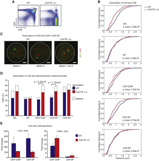
T cell fate is associated with mutually exclusive expression of CD4 or CD8 in helper and cytotoxic T cells, respectively. How expression of one locus is temporally coordinated with repression of the other has been a long-standing enigma, though we know RUNX transcription factors activate the Cd8 locus, silence the Cd4 locus, and repress the Zbtb7b locus (encoding the transcription factor ThPOK), which is required for CD4 expression. Here we found that nuclear organization was altered by interplay among members of this transcription factor circuitry: RUNX binding mediated association of Cd4 and Cd8 whereas ThPOK binding kept the loci apart. Moreover, targeted deletions within Cd4 modulated CD8 expression and pericentromeric repositioning of Cd8. Communication between Cd4 and Cd8 thus appears to enable long-range epigenetic regulation to ensure that expression of one excludes the other in mature CD4 or CD8 single-positive (SP) cells.


Figures

None
Graphical abstract
Figure 1
Figure 1
Cd4 and Cd8 Associate in Murine CD8-Expressing Cells (A) Confocal microscopy sections showing a range of distances between the Cd4 and Cd8 loci. Scale bars represent 1 μm. (B) Association of Cd4-Cd8 in DP T cells compared to B cells (top); association of Cd4-Cd8 compared to Tcrb-Lrig1 in DP T cells (bottom). The separation of signals is plotted as a cumulative frequency of association. Association of Cd4-Cd8 in DP cells was increased compared to B cells and higher than association of Tcrb-Lrig1 in DP cells. The Kolmogorov-Smirnov test was used for statistical analysis. At least three independent experiments were performed for each data set. n = 238–264 alleles. (C) Association of Cd4-Cd8 in developing T cell populations and statistical analysis between specified stages. Association is increased in DP and CD8 SP cells. n = 166–238 alleles. (D) Images of Cd4 and Cd8 and their individual chromosome 6 territories in DP cells. See also Figure S1 and Table S1.
Figure 2
Figure 2
The E8I and E8II Enhancers Promote Cd8 Transcription and Cd4-Cd8 Association (A) Cd4-Cd8 association in wild-type and E8I-deficient CD8 SP cells (E8I Δ/Δ). Association is decreased in E8I-deficient CD8 SP compared to wild-type cells. n = 230–264 alleles. (B) Cd8a RNA expression in wild-type and E8I-deficient DP and CD8 SP cells. Standard error bars were calculated from three independent experiments. (C) Flow cytometry analysis of wild-type and E8IE8II double-mutant DP cells (E8I Δ/Δ E8II Δ/Δ) (TCRβintCD24+). (D) Cd4-Cd8 association in wild-type and E8IE8II double-mutant DP cells. Statistical analyses are between specified genotypes. Association is lower in CD8+ and CD8lo E8IE8II double-mutant than in wild-type cells. Confocal microscopy sections of Cd4-Cd8 distances are representative of each genotype. Scale bars represent 1 μm. n = 204–248 alleles. (E) RT-PCR analysis of Cd8a (top) or Cd4 (bottom) expression in wild-type and E8IE8II double-mutant DP cells. Standard error bars were calculated from two independent experiments. (F) Cd4 or Cd8 association with pericentromeric heterochromatin in wild-type and E8IE8II double-mutant DP cells. Cd8 recruitment is higher in double-mutant than in wild-type control cells. See also Figure S2 and Table S2.
Figure 3
Figure 3
Cd4-Cd8 Association Requires the RUNX Binding Partner CBFβ (A) Flow cytometry analysis of wild-type and CBFβ-deficient thymocytes (conditional CbfbF/F crossed to Lck-cre). (B) Cd4-Cd8 association in wild-type and CBFβ-deficient DN and DP T cells, including statistical analysis. Association is lower in CBFβ-deficient DP cells than in wild-type counterparts. n = 228–264 alleles. (C) Confocal microscopy sections of Cd4-Cd8 distances representative of wild-type or CBFβ-deficient DP cells. Scale bars represent 1 μm. See also Figure S3.
Figure 4
Figure 4
ThPOK Inhibits Cd4-Cd8 Association (A) Cd8a RNA expression in peripheral CD8+ T cells transduced with empty pMIGR, pMIGR.ThPOK, or pMIGR.ThPOK.HD. (B) Flow cytometry analysis of wild-type and Zbtb7bhd/hd mature SP cells. (C) RUNX3 staining in wild-type CD8 cells and wild-type or Zbtb7bhd/hd CD4 SP cells. Scale bars represent 1 μm. (D) Cd4-Cd8 association in wild-type and Zbtb7bhd/hd CD4 SP cells. Association is higher in Zbtb7bhd/hd CD4 SP than in wild-type cells. n = 206–218 alleles. (E) Cd4 recruitment to pericentromeric heterochromatin in wild-type and Zbtb7bhd/hd CD4 SP cells. Recruitment is higher in Zbtb7bhd/hd cells. (F) RT-PCR analysis of Cd4 or Cd8a expression in wild-type and Zbtb7bhd/hd CD4 SP cells. Standard error bars were calculated from three independent experiments. (G) Cd4-Cd8 association in wild-type and ThPOK transgenic DP cells. Cd4-Cd8 association is lower in ThPOK transgenic DP cells. n = 316–356 alleles. See also Figure S4 and Table S3.
Figure 5
Figure 5
The Cd4 Proximal Enhancer Inhibits Cd4-Cd8 Association (A) Flow cytometry analysis of wild-type and Cd4 PE-deficient thymocytes (Cd4 PE Δ/Δ). (B) Cd4-Cd8 association in wild-type and Cd4 PE-deficient cells, including statistical analysis. Association is higher in Cd4 PE-deficient DN, CD4+CD8lo, and CD4 SP cells than in wild-type cells. n = 196–286 alleles. (C) Confocal microscopy sections of Cd4-Cd8 distances representative of each genotype. Scale bars represent 1 μm. (D) Recruitment of Cd4 to pericentromeric heterochromatin in wild-type and Cd4 PE-deficient cells. Recruitment is higher in Cd4 PE-deficient DN, CD4+CD8lo and CD4 SP than in wild-type cells. (E) RT-PCR analysis of Cd4 or Cd8a expression in wild-type and Cd4 PE-deficient cells. Standard error bars were calculated from three independent experiments. See also Figure S5 and Table S4.
Figure 6
Figure 6
The Cd4 Silencer Mediates Cd4-Cd8 Association (A) Flow cytometry analysis of wild-type and Cd4 sil-deficient thymocytes (Cd4 sil Δ/Δ). (B) Cd4-Cd8 association in wild-type and Cd4 sil-deficient cells, including statistical analysis. Association is lower in Cd4 sil-deficient DP, CD4+CD8lo, and CD8 SP than in wild-type cells. n = 210–340 alleles. (C) Confocal microscopy sections of Cd4-Cd8 distances representative of each genotype. Scale bars represent 1 μm. (D) Cd8 recruitment to pericentromeric heterochromatin in wild-type and Cd4 sil-deficient cells. Recruitment is higher in Cd4 sil-deficient DP and CD8 SP cells than in wild-type cells. See also Figure S6 and Table S5.
Figure 7
Figure 7
CD4 and CD8 Also Associate in Human CD8-Expressing Cells (A) 3D DNA FISH on human sorted CD8+ T cells, CD4+ T cells, and B cells. (B) CD4-CD8 association in B and T cells, including statistical analysis between specified cell types. Association in CD8+ T cells is increased compared to CD4+ T and B cells. n = 196–206 alleles. See also Figure S7.

References

    1. Bacher C.P., Guggiari M., Brors B., Augui S., Clerc P., Avner P., Eils R., Heard E. Transient colocalization of X-inactivation centres accompanies the initiation of X inactivation. Nat. Cell Biol. 2006;8:293–299. - PubMed
    1. Bilic I., Koesters C., Unger B., Sekimata M., Hertweck A., Maschek R., Wilson C.B., Ellmeier W. Negative regulation of CD8 expression via Cd8 enhancer-mediated recruitment of the zinc finger protein MAZR. Nat. Immunol. 2006;7:392–400. - PMC - PubMed
    1. Brown K.E., Baxter J., Graf D., Merkenschlager M., Fisher A.G. Dynamic repositioning of genes in the nucleus of lymphocytes preparing for cell division. Mol. Cell. 1999;3:207–217. - PubMed
    1. Campbell R. Cambridge University Press; Cambridge, UK: 1989. Statistics for Biologists.
    1. Chambeyron S., Bickmore W.A. Chromatin decondensation and nuclear reorganization of the HoxB locus upon induction of transcription. Genes Dev. 2004;18:1119–1130. - PMC - PubMed

Publication types

MeSH terms

Substances