

Case Reports
doi: 10.1102/1470-7330.2011.0005.
Omental inflammatory myofibroblastic tumour mimicking peritoneal carcinomatosis
Affiliations
- PMID: 21435987
- PMCID: PMC3080123
- DOI: 10.1102/1470-7330.2011.0005
Item in Clipboard
Case Reports
Omental inflammatory myofibroblastic tumour mimicking peritoneal carcinomatosis
Cancer Imaging.
.
Abstract
Inflammatory myofibroblastic tumour (IMFT) is a relatively uncommon neoplasm with unpredictable malignant potential known to occur anywhere in the body. IMFT involving the omentum is a very rare entity with less than 15 cases reported so far. We report a case of omental IMFT in a 15-year-old girl who presented with multiple peritoneal masses on imaging and the diagnosis was confirmed on histopathology. In addition to its uncommon location, its presentation as multiple masses is extremely uncommon. This uncommon presentation as multifocal masses needs to be distinguished from other causes of peritoneal carcinomatosis.
Figures
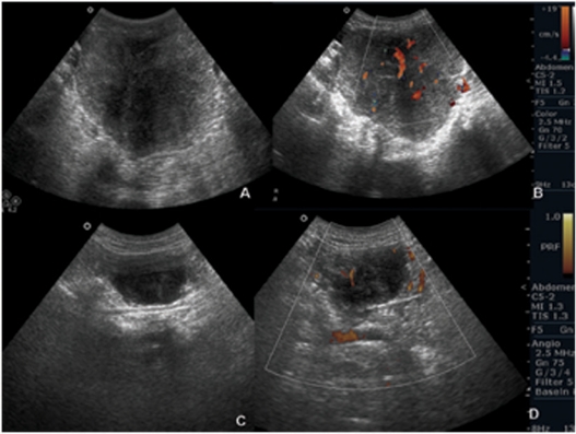
US images show heterogeneous hypoechoic lesions in the pelvis (a) and on the parietal peritoneum (c). Note internal vascularity on colour Doppler (b,d).
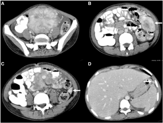
Contrast-enhanced CT images show a lobulated hypervascular lesion in the pelvis with free fluid (a). Note multiple peritoneal-based lesions (arrows) (b–d).
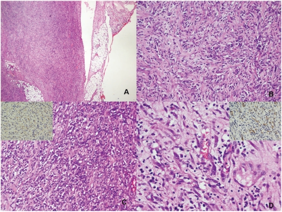
(a) The periphery of the tumour with infiltration into fat (H&E); (b) spindle-shaped tumour cells in short interlacing fascicles admixed with moderate chronic inflammatory cells rich in plasma cells (H&E); (c) spindle cell populations with plasma cell infiltrate with the tumour cells showing cytoplasmic ALK positivity (inset: ALK immunostaining); and (d) some tumour cells are elongated with abundant eosinophilic cytoplasm (H&E) showing SMA positivity (inset: SMA immunostaining) indicating the myofibroblastic nature.
References
Publication types
MeSH terms
LinkOut - more resources
Full Text Sources
Medical