. 2011 Apr 5;108(14):5620-5.
doi: 10.1073/pnas.1014890108. Epub 2011 Mar 21.

Histone deacetylase inhibitor treatment dramatically reduces cholesterol accumulation in Niemann-Pick type C1 mutant human fibroblasts

Affiliations

Histone deacetylase inhibitor treatment dramatically reduces cholesterol accumulation in Niemann-Pick type C1 mutant human fibroblasts

Nina H Pipalia et al. Proc Natl Acad Sci U S A. .

Abstract

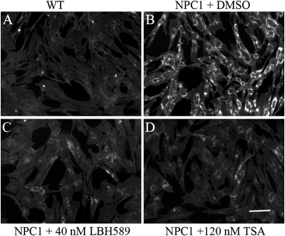
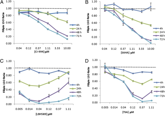
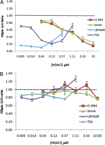
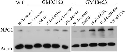
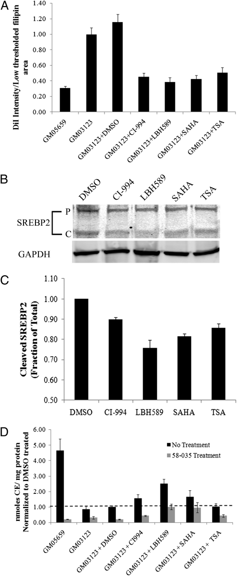
Niemann-Pick type C (NPC) disease is predominantly caused by mutations in the NPC1 protein that affect intracellular cholesterol trafficking and cause accumulation of unesterified cholesterol and other lipids in lysosomal storage organelles. We report the use of a series of small molecule histone deacetylase (HDAC) inhibitors in tissue culture models of NPC human fibroblasts. Some HDAC inhibitors lead to a dramatic correction in the NPC phenotype in cells with either one or two copies of the NPC1(I1061T) mutation, and for several of the inhibitors, correction is associated with increased expression of NPC1 protein. Increased NPC1(I1061T) protein levels may partially account for the correction of the phenotype, because this mutant can promote cholesterol efflux if it is delivered to late endosomes and lysosomes. The HDAC inhibitor treatment is ineffective in an NPC2 mutant human fibroblast line. Analysis of the isoform selectivity of the compounds used implicates HDAC1 and/or HDAC2 as likely targets for the observed correction, although other HDACs may also play a role. LBH589 (panobinostat) is an orally available HDAC inhibitor that crosses the blood-brain barrier and is currently in phase III clinical trials for several types of cancer. It restores cholesterol homeostasis in cultured NPC1 mutant fibroblasts to almost normal levels within 72 h when used at 40 nM. The findings that HDAC inhibitors can correct cholesterol storage defects in human NPC1 mutant cells provide the potential basis for treatment options for NPC disease.


Conflict of interest statement

The authors declare no conflict of interest.

Figures

Fig. 1.
Fig. 1.
Representative filipin images of HDACi-treated NPC1 mutant fibroblasts. NPC1 mutant human fibroblasts GM03123 were treated with HDACi LBH589 or 120 TSA and incubated for 48 h. Cells were subsequently fixed, stained with filipin, and imaged by epifluorescence microscopy at 10× magnification. (A) WT human fibroblasts GM05659. (B) NPC1 human fibroblasts GM03123 treated with DMSO. (C) GM03123 cells after 48-h treatment with 40 nM LBH589. (D) GM03123 cells after 48-h treatment with 120 nM TSA. (Scale bar, 25 μM.)
Fig. 2.
Fig. 2.
Dose- and time-dependence plots for HDACi (set 1). NPC1 mutant human fibroblasts GM03123 were treated with (A) CI-994, (B) SAHA, (C) LBH589, or (D) TSA at various concentrations and for varying time followed by fixing, staining with filipin, and imaging. DMSO was used as a solvent control. Images were analyzed to obtain filipin fluorescence power in bright spots (filipin LSO ratio), and data were normalized to corresponding DMSO-treated cells. Data shown are averages of three independent experiments totaling 60 images (five wells × four sites × three experiments). The dotted horizontal lines indicate mean values for the solvent control; error bars represent SE.
Fig. 3.
Fig. 3.
Dose dependence for NPC1 mutant cells GM18453 and NPC2 mutant cells GM18445 in the presence of HDACi. NPC1 mutant human fibroblasts GM18453 (A) and NPC2 mutant human fibroblasts GM18445 (B) were treated with CI-994, SAHA, LBH589, or TSA at various concentrations for 48 h followed by fixing, staining with filipin, and imaging. DMSO was used as a solvent control. Images were analyzed to obtain filipin LSO ratio, and data were normalized to corresponding DMSO-treated cells. Data shown are averages of three independent experiments totaling 60 images (five wells × four sites × three experiments). The dotted horizontal lines indicate mean values for the solvent control; error bars represent SE.
Fig. 4.
Fig. 4.
Effect of HDACi on NPC1 expression. WT human fibroblast GM05659 and NPC1 mutant human fibroblasts GM03123 and GM18453 were treated with LBH589, TSA, or SAHA. As a control, cells were treated with DMSO. After 48 h, Western blot analysis was performed. The membrane was probed with mouse monoclonal anti-human NPC1 antibody, and anti–α-actin was used as a loading control.
Fig. 5.
Fig. 5.
Characterization of HDACi. (A) DiI-LDL uptake. NPC1 mutant GM03123 fibroblasts were treated with HDACi as described in Materials and Methods. DiI intensity per cell area was measured after treatment with 10 μM CI-994, 0.04 μM LBH589, 10 μM SAHA, or 0.37 μM TSA. DMSO treatment was used as a vehicle control, and WT GM05659 fibroblasts were used as a reference. Data shown are from three independent experiments, and 20 images were acquired per experiment. Error bar represents SE, and P values compared with DMSO control were <0.001 for all treatments (Student t test). (B) SREBP2 processing. GM03123 cells were treated with 10 μM CI-994, 0.04 μM LBH589, 10 μM SAHA, or 0.37 μM TSA. As a control, cells were treated with DMSO. After 18 h, Western blot analysis was performed. The membrane was probed with mouse monoclonal anti-SREBP2 antibody. (C) Quantification of SREBP2. The ratio of cleaved SREBP2 to total SREBP2 (precursor + cleaved form) is plotted for treatments with HDACi, using DMSO treatment as a control. Error bar represents SE, and P values compared with DMSO control were <0.05 for all treatments (Student t test). (D) ACAT assay. WT and NPC1 mutant GM05659 and GM03123 fibroblasts were plated in growth medium with 10% FBS at 37 °C. ACAT assay was performed as described in Materials and Methods. The nanomoles of cholesteryl-[14C]-oleate per milligram cell protein were calculated. Data for HDACi-treated cells were normalized to DMSO-treated controls and are presented as mean ± SE. The LBH589-treated cells had a statistically significant (P < 0.05) increase in esterification in the absence of the ACAT inhibitor (Student Newman–Kuels multiple comparison). Two independent experiments were conducted, and each experiment had three data points.

References

    1. Shahbazian MD, Grunstein M. Functions of site-specific histone acetylation and deacetylation. Annu Rev Biochem. 2007;76:75–100. - PubMed
    1. Budillon A, Bruzzese F, Di Gennaro E, Caraglia M. Multiple-target drugs: Inhibitors of heat shock protein 90 and of histone deacetylase. Curr Drug Targets. 2005;6:337–351. - PubMed
    1. de Ruijter AJ, van Gennip AH, Caron HN, Kemp S, van Kuilenburg AB. Histone deacetylases (HDACs): Characterization of the classical HDAC family. Biochem J. 2003;370:737–749. - PMC - PubMed
    1. Gregoretti IV, Lee YM, Goodson HV. Molecular evolution of the histone deacetylase family: Functional implications of phylogenetic analysis. J Mol Biol. 2004;338:17–31. - PubMed
    1. Dokmanovic M, Clarke C, Marks PA. Histone deacetylase inhibitors: Overview and perspectives. Mol Cancer Res. 2007;5:981–989. - PubMed

Publication types

MeSH terms