. 2011 Apr 5;108(14):5867-72.
doi: 10.1073/pnas.1017975108. Epub 2011 Mar 21.

Agonist activation of alpha7 nicotinic acetylcholine receptors via an allosteric transmembrane site

Affiliations

Agonist activation of alpha7 nicotinic acetylcholine receptors via an allosteric transmembrane site

Jaskiran K Gill et al. Proc Natl Acad Sci U S A. .

Abstract

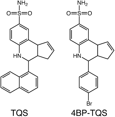
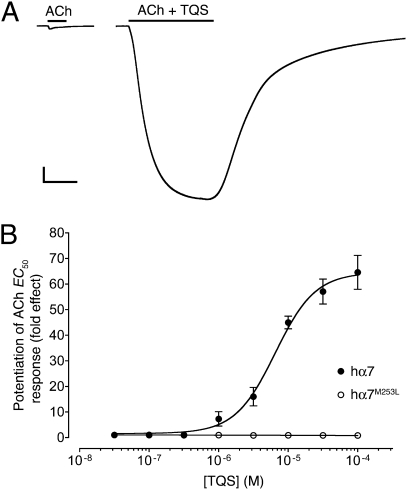
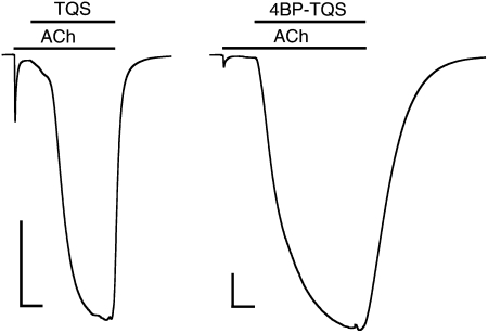
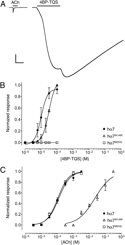
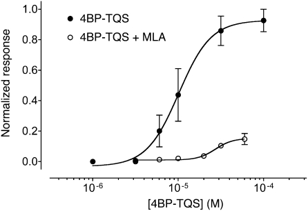
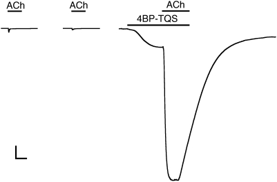
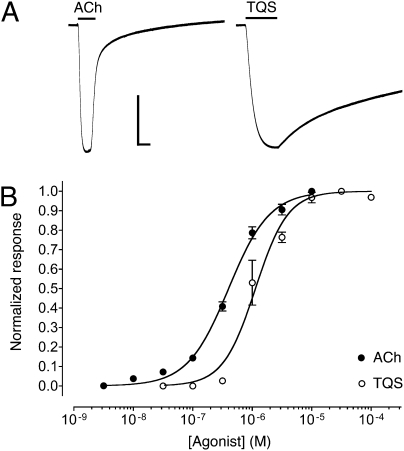
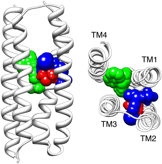
Conventional nicotinic acetylcholine receptor (nAChR) agonists, such as acetylcholine, act at an extracellular "orthosteric" binding site located at the interface between two adjacent subunits. Here, we present evidence of potent activation of α7 nAChRs via an allosteric transmembrane site. Previous studies have identified a series of nAChR-positive allosteric modulators (PAMs) that lack agonist activity but are able to potentiate responses to orthosteric agonists, such as acetylcholine. It has been shown, for example, that TQS acts as a conventional α7 nAChR PAM. In contrast, we have found that a compound with close chemical similarity to TQS (4BP-TQS) is a potent allosteric agonist of α7 nAChRs. Whereas the α7 nAChR antagonist metyllycaconitine acts competitively with conventional nicotinic agonists, metyllycaconitine is a noncompetitive antagonist of 4BP-TQS. Mutation of an amino acid (M253L), located in a transmembrane cavity that has been proposed as being the binding site for PAMs, completely blocks agonist activation by 4BP-TQS. In contrast, this mutation had no significant effect on agonist activation by acetylcholine. Conversely, mutation of an amino acid located within the known orthosteric binding site (W148F) has a profound effect on agonist potency of acetylcholine (resulting in a shift of ∼200-fold in the acetylcholine dose-response curve), but had little effect on the agonist dose-response curve for 4BP-TQS. Computer docking studies with an α7 homology model provides evidence that both TQS and 4BP-TQS bind within an intrasubunit transmembrane cavity. Taken together, these findings provide evidence that agonist activation of nAChRs can occur via an allosteric transmembrane site.


Conflict of interest statement

The authors declare no conflict of interest.

Figures

Fig. 1.
Fig. 1.
Chemical structure of TQS and 4BP-TQS.
Fig. 2.
Fig. 2.
Positive allosteric modulation of α7 nAChRs by TQS, examined by two-electrode voltage-clamp recording in Xenopus oocytes. (A) Representative recordings are shown illustrating responses to the application of acetylcholine (100 μM) (Left) and of acetylcholine (100 μM) coapplied with TQS (100 μM) (Right). The duration of agonist applications are indicated by a horizontal line. (Scale bars: vertical, 1 μA; horizontal, 5 s.) (B) Dose-response data are presented for a range of concentrations of TQS (0.03–100 μM) on responses evoked by a submaximal (EC50) concentration of acetylcholine. Data were obtained with either wild-type α7 nAChRs (●) or α7 nAChRs containing the M253L mutation (○). Data are means ± SEM of at least three independent experiments, each from different oocytes.
Fig. 3.
Fig. 3.
TQS and 4BP-TQS facilitate recovery of α7 nAChRs from desensitization. Prolonged exposure of α7 nAChRs to a high concentration of acetylcholine (100 μM) results in receptor activation, followed by rapid desensitization. In the continued presence of acetylcholine (100 μM), coapplication of either TQS (10 μM) (Left) or 4BP-TQS (10 μM) (Right) results in reactivation of desensitized receptors. Applications of acetylcholine and allosteric modulators (TSQ or 4BP-TQS) are indicated by horizontal lines. (Scale bars: vertical, 0.5 μA; horizontal, 10 s.)
Fig. 4.
Fig. 4.
Agonist activation of α7 nAChRs by acetylcholine and 4BP-TQS, examined by two-electrode voltage-clamp recordings in Xenopus oocytes. (A) Representative recordings are shown illustrating responses to the application of acetylcholine (3 mM) (Left) and of 4BP-TQS (60 μM) (Right). The duration of agonist applications are indicated by a horizontal line. (Scale bars: vertical, 1 μA; horizontal, 5 s.) Dose-response data are presented for a range of concentrations of 4BP-TQS (B) or acetylcholine (C), with wild-type α7 nAChRs (●) or with α7 nAChRs containing either the W148F mutation (△) or the M253L mutation (□). Data are means ± SEM of at least three independent experiments, each from different oocytes.
Fig. 5.
Fig. 5.
MLA is a noncompetitive antagonist of 4BP-TQS. Dose-response data are presented for a range of concentrations of acetylcholine acting on α7 nAChRs either in the absence (●) or presence (○) of MLA (5 nM). In all cases, MLA was preapplied for 15 s and then coapplied with 4BP-TQS. Data are means ± SEM of at least three independent experiments, each from different oocytes.
Fig. 6.
Fig. 6.
4BP-TQS acts as potentiator of acetylcholine responses. Representative traces showing agonist responses on α7 nAChRs to a maximal concentration (3 mM) (Left) and an EC10 concentration (25 μM) (Center) of acetylcholine. Also shown (Right) is a response to 4BP-TQS (10 μM). After the response to 4BP-TQS had reached a plateau, acetylcholine (25 μM) was coapplied with 4BP-TQS (10 μM), resulting in a secondary response of much greater magnitude (542 ± 32; n = 3) than was observed when the same concentration of acetylcholine was applied alone (Center).
Fig. 7.
Fig. 7.
The α7 nAChR transmembrane mutation L247T converts TQS from a PAM into an agonist. (A) Representative recordings are shown illustrating responses to the application of acetylcholine (3 μM) (Left) and of TQS (100 μM) (Right). The duration of agonist applications are indicated by a horizontal line. (Scale bars: vertical, 0.5 μA; horizontal, 5 s.) (B) Dose-response data are presented for a range of concentrations of acetylcholine (●) and TQS (○) acting on α7 nAChRs containing the transmembrane L247T mutation. Data are means ± SEM of at least three independent experiments, each from different oocytes.
Fig. 8.
Fig. 8.
Computer-docking simulation performed with a homology model of the α7 transmembrane domain (21). The backbone of the four transmembrane α-helices (TM1–TM4) are shown in gray. The side chain of M253 is in red (with atoms represented as spheres) the lowest energy (highest predicted binding affinity) docked position of 4BP-TQS is illustrated in blue. For comparison, the lowest energy docked position of TQS is illustrated in green. The model is shown from a side-on view (Left) and as viewed from above, looking down from the extracellular face of the lipid membrane (Right).

References

    1. Lester HA, Dibas MI, Dahan DS, Leite JF, Dougherty DA. Cys-loop receptors: New twists and turns. Trends Neurosci. 2004;27:329–336. - PubMed
    1. Le Novère N, Changeux J-P. Molecular evolution of the nicotinic acetylcholine receptor: An example of multigene family in excitable cells. J Mol Evol. 1995;40:155–172. - PubMed
    1. Millar NS, Gotti C. Diversity of vertebrate nicotinic acetylcholine receptors. Neuropharmacology. 2009;56:237–246. - PubMed
    1. Changeux J-P, Taly A. Nicotinic receptors, allosteric proteins and medicine. Trends Mol Med. 2008;14:93–102. - PubMed
    1. Weiland S, Bertrand D, Leonard S. Neuronal nicotinic acetylcholine receptors: From the gene to the disease. Behav Brain Res. 2000;113:43–56. - PubMed

Publication types

MeSH terms

LinkOut - more resources