

Structure and binding analysis of Polyporus squamosus lectin in complex with the Neu5Ac{alpha}2-6Gal{beta}1-4GlcNAc human-type influenza receptor
- PMID: 21436237
- PMCID: PMC3110490
- DOI: 10.1093/glycob/cwr030
Structure and binding analysis of Polyporus squamosus lectin in complex with the Neu5Ac{alpha}2-6Gal{beta}1-4GlcNAc human-type influenza receptor
Abstract
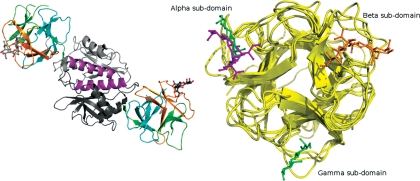
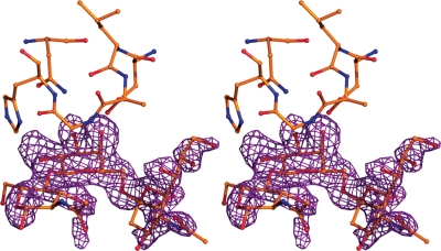
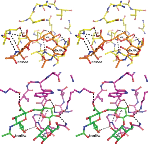
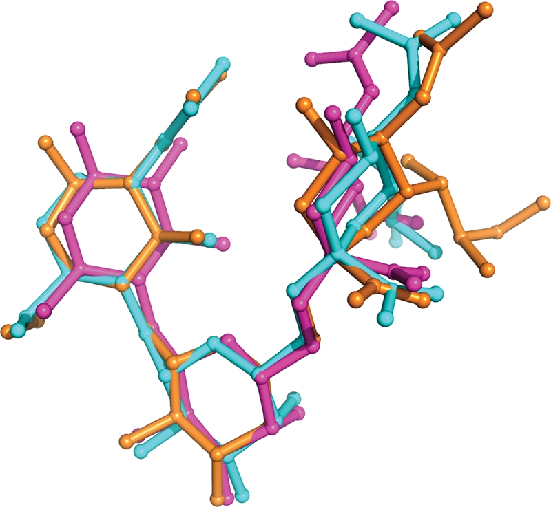
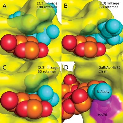
Glycan chains that terminate in sialic acid (Neu5Ac) are frequently the receptors targeted by pathogens for initial adhesion. Carbohydrate-binding proteins (lectins) with specificity for Neu5Ac are particularly useful in the detection and isolation of sialylated glycoconjugates, such as those associated with pathogen adhesion as well as those characteristic of several diseases including cancer. Structural studies of lectins are essential in order to understand the origin of their specificity, which is particularly important when employing such reagents as diagnostic tools. Here, we report a crystallographic and molecular dynamics (MD) analysis of a lectin from Polyporus squamosus (PSL) that is specific for glycans terminating with the sequence Neu5Acα2-6Galβ. Because of its importance as a histological reagent, the PSL structure was solved (to 1.7 Å) in complex with a trisaccharide, whose sequence (Neu5Acα2-6Galβ1-4GlcNAc) is exploited by influenza A hemagglutinin for viral adhesion to human tissue. The structural data illuminate the origin of the high specificity of PSL for the Neu5Acα2-6Gal sequence. Theoretical binding free energies derived from the MD data confirm the key interactions identified crystallographically and provide additional insight into the relative contributions from each amino acid, as well as estimates of the importance of entropic and enthalpic contributions to binding.
Figures
Similar articles
-
Cloning, expression in Escherichia coli and characterization of the recombinant Neu5Acalpha2,6Galbeta1,4GlcNAc-specific high-affinity lectin and its mutants from the mushroom Polyporus squamosus.Biochem J. 2004 Sep 1;382(Pt 2):667-75. doi: 10.1042/BJ20040391. Biochem J. 2004. PMID: 15176950 Free PMC article.
-
Mistletoe lectin I is a sialic acid-specific lectin with strict preference to gangliosides and glycoproteins with terminal Neu5Ac alpha 2-6Gal beta 1-4GlcNAc residues.Biochemistry. 2004 Mar 23;43(11):2996-3007. doi: 10.1021/bi0301892. Biochemistry. 2004. PMID: 15023051
-
Distribution of sialoglycoconjugates in the oviductal isthmus of the horse during anoestrus, oestrus and pregnancy: a lectin histochemistry study.Eur J Histochem. 2004 Oct-Dec;48(4):403-12. doi: 10.4081/914. Eur J Histochem. 2004. PMID: 15718207
-
Glycans as receptors for influenza pathogenesis.Glycoconj J. 2010 Aug;27(6):561-70. doi: 10.1007/s10719-010-9303-4. Epub 2010 Aug 24. Glycoconj J. 2010. PMID: 20734133 Free PMC article. Review.
-
I-type lectins.Biochim Biophys Acta. 2002 Sep 19;1572(2-3):294-316. doi: 10.1016/s0304-4165(02)00316-1. Biochim Biophys Acta. 2002. PMID: 12223277 Review.
Cited by
-
Genetically encoded fragment-based discovery of glycopeptide ligands for carbohydrate-binding proteins.J Am Chem Soc. 2015 Apr 29;137(16):5248-51. doi: 10.1021/ja511237n. Epub 2015 Apr 16. J Am Chem Soc. 2015. PMID: 25860443 Free PMC article.
-
Hitting the sweet spot-glycans as targets of fungal defense effector proteins.Molecules. 2015 May 6;20(5):8144-67. doi: 10.3390/molecules20058144. Molecules. 2015. PMID: 25955890 Free PMC article. Review.
-
Nuclear Magnetic Resonance and Molecular Dynamics Simulation of the Interaction between Recognition Protein H7 of the Novel Influenza Virus H7N9 and Glycan Cell Surface Receptors.Biochemistry. 2016 Dec 6;55(48):6605-6616. doi: 10.1021/acs.biochem.6b00693. Epub 2016 Nov 23. Biochemistry. 2016. PMID: 27933797 Free PMC article.
-
Enzymatic basis for N-glycan sialylation: structure of rat α2,6-sialyltransferase (ST6GAL1) reveals conserved and unique features for glycan sialylation.J Biol Chem. 2013 Nov 29;288(48):34680-98. doi: 10.1074/jbc.M113.519041. Epub 2013 Oct 23. J Biol Chem. 2013. PMID: 24155237 Free PMC article.
-
Human osteoarthritic knee cartilage: fingerprinting of adhesion/growth-regulatory galectins in vitro and in situ indicates differential upregulation in severe degeneration.Histochem Cell Biol. 2014 Oct;142(4):373-88. doi: 10.1007/s00418-014-1234-x. Epub 2014 Jul 1. Histochem Cell Biol. 2014. PMID: 24981556
References
-
- Adams PD, Grosse-Kunstleve RW, Hung LW, Ioerger TR, McCoy AJ, Moriarty NW, Read RJ, Sacchettini JC, Sauter NK, Terwilliger TC. PHENIX: Building new software for automated crystallographic structure determination. Acta Crystallogr Sect D Biol Crystallogr. 2002;58:1948–1954. doi:10.1107/S0907444902016657. - DOI - PubMed
-
- Angata T, Varki A. Chemical diversity in the sialic acids and related α-keto acids: An evolutionary perspective. Chem Rev. 2002;102:439–469. doi:10.1021/cr000407m. - DOI - PubMed
-
- Baum LG, Paulson JC. Sialyloligosaccharides of the respiratory epithelium in the selection of human influenza virus receptor specificity. Acta Histochem Suppl. 1990;40:35–38. - PubMed
-
- Benallal M, Zotter H, Anner RM, Lacotte D, Moosmayer M, Anner BM. Maackia amurensis agglutinin discriminates between normal and chronic leukemic human lymphocytes. Biochem Biophys Res Commun. 1995;209:921–929. doi:10.1006/bbrc.1995.1586. - DOI - PubMed
-
- Bernstein FC, Koetzle TF, Williams GJ, Meyer EF, Jr, Brice MD, Rodgers JR, Kennard O, Shimanouchi T, Tasumi M. The Protein Data Bank: A computer-based archival file for macromolecular structures. J Mol Biol. 1977;112:535–542. doi:10.1016/S0022-2836(77)80200-3. - DOI - PubMed
Publication types
MeSH terms
Substances
Grants and funding
LinkOut - more resources
Full Text Sources
Other Literature Sources
