Comparative Study
. 2011 Jul 1;52(7):4703-9.
doi: 10.1167/iovs.10-7077.

An ENU-induced mutation in the Mertk gene (Mertknmf12) leads to a slow form of retinal degeneration

Affiliations
Comparative Study

An ENU-induced mutation in the Mertk gene (Mertknmf12) leads to a slow form of retinal degeneration

Dennis M Maddox et al. Invest Ophthalmol Vis Sci. .

Abstract

Purpose: To determine the basis and to characterize the phenotype of a chemically induced mutation in a mouse model of retinal degeneration.

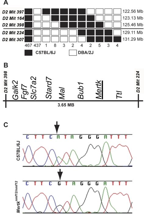
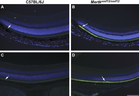
Methods: Screening by indirect ophthalmoscopy identified a line of N-ethyl-N-nitrosourea (ENU) mutagenized mice demonstrating retinal patches. Longitudinal studies of retinal histologic sections showed photoreceptors in the peripheral retina undergoing slow, progressive degeneration. The mutation was named neuroscience mutagenesis facility 12 (nmf12), and mapping localized the critical region to Chromosome 2.

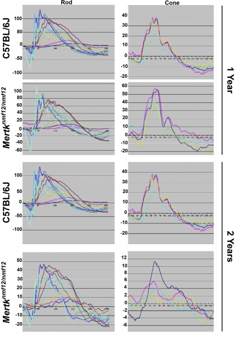
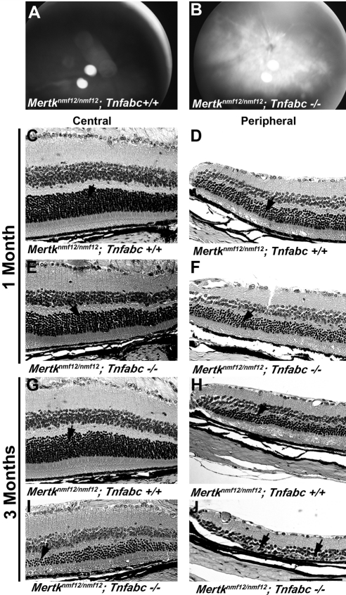
Results: Sequencing of nmf12 DNA revealed a point mutation in the c-mer tyrosine kinase gene, designated Mertk(nmf12). We detected elevated levels of tumor necrosis factor (Tnf, previously Tnfa) in retinas of Mertk(nmf12) homozygotes relative to wild-type controls and investigated whether the increase of TNF, an inflammatory cytokine produced by macrophages/monocytes that signals intracellularly to cause necrosis or apoptosis, could underlie the retinal degeneration observed in Mertk(nmf12) homozygotes. Mertk(nmf12) homozygous mice were mated to mice lacking the entire Tnf gene and partial coding sequences of the Lta (Tnfb) and Ltb (Tnfc) genes.(2) B6.129P2-Ltb/Tnf/Lta(tm1Dvk)/J homozygotes did not exhibit a retinal degeneration phenotype and will, hereafter, be referred to as Tnfabc(-/-) mice. Surprisingly, mice homozygous for both the Mertk(nmf12) and the Ltb/Tnf/Lta(tm1Dvk) allele (Tnfabc(-/-)) demonstrated an increase in the rate of retinal degeneration.

Conclusions: These findings illustrate that a mutation in the Mertk gene leads to a significantly slower progressive retinal degeneration compared with other alleles of Mertk. These results demonstrate that TNF family members play a role in protecting photoreceptors of Mertk(nmf12) homozygotes from cell death.


Figures

Figure 1.
Figure 1.
Mapping and cloning of the nmf12 mutation. (A) A haplotype map is shown, representing the location and number of crossovers in 936 meioses observed in 468 affected and unaffected progeny-tested mice. The numbers at the bottom of the graph represent the number of chromosomes bearing each depicted crossover. The critical interval spans 3.65 Mb between markers D2Mit398 (125.46 Mb) and D2Mit224 (129.11 Mb). Within this critical region (B) lies Mertk, a gene in which mutations have been shown to cause retinal degeneration. (C) Sequence chromatograms from control, C57BL/6J, and Mertknmf12 homozygotes. Analysis of these sequences reveals an A-to-G transition at base pair 2237 (A2237G).
Figure 2.
Figure 2.
Complementation testing between Mertknmf12 and Mertktm1Gkm. Indirect ophthalmoscopy shows that at P45, the retinas of Mertknmf12/Mertktm1Gkm compound heterozygous mice demonstrate pan-retinal fundus patches (B), whereas the Mertknmf12/+ heterozygous littermates (as well as Mertknmf12 homozygotes) demonstrate a normal appearance in the central retina (A). Histologic examination of central and peripheral sections from the Mertknmf12/+ heterozygous littermates (C and E, respectively) appears normal. In contrast, the central retina of Mertknmf12/tm1Gkm compound heterozygotes (D) demonstrates slight thinning of the ONL (arrows), whereas the peripheral retina (F) demonstrates severe photoreceptor loss (arrows). Failure of the Mertknmf12/Mertktm1Gkm compound heterozygous mice to show a normal phenotype provides strong evidence that Mertknmf12 and Mertktm1Gkm do not complement and are likely to be allelic mutations. Scale bar, 50 μm (CF).
Figure 3.
Figure 3.
Analysis of Mertknmf12 protein expression and function. (A) Depiction of protein alignment between C57BL/6J and Mertknmf12 homozygotes (amino acids presented in randomly selected colors). A substitution of arginine for histidine is evident at amino acid 716 (H716R; arrow). This residue is conserved across species (B). (C) Schematic showing that the mutated amino acid falls within the tyrosine kinase domain of the Mertk gene. Western blot analysis shows a 2.5-fold reduction of MERTK protein in Mertknmf12 homozygous retinas (D). The blot was stripped and probed again with an antibody against β-actin as a loading control (E).
Figure 4.
Figure 4.
Analysis of the Mertknmf12 retinal phenotype. Fundus photographs of 3-month-old C57BL/6J (A) or homozygous Mertknmf12 (B) mice. Note the abnormal whitening of the peripheral (P) and central (C) retina that is likely to correspond to areas of ONL degeneration. Histologic sections of the peripheral retina from a P30 C57BL/6J mouse (C). The ONL in retinas of Mertknmf12 homozygotes aged P30, P45, and 6 months are shown (DF, respectively). (D, arrow) Beginning of ONL cell thinning. This ONL thinning progresses over time. (E, F, black arrows) Thinning ONL. Pyknotic nuclei are present (E, F, white arrows) suggesting that cell death is taking place. Few TUNEL-positive cells (white arrow) are present in the retinas of P45 C57BL/6J mice (G), whereas many TUNEL-positive cells (white arrows) are present throughout the ONL in retinas of Mertknmf12 homozygotes (H). Scale bar, 50 μm (CH).
Figure 5.
Figure 5.
Analysis of the ERG function in eyes of Mertknmf12 mutants. Although dying cells are present throughout the retinas of Mertknmf12 homozygotes, the majority of photoreceptor cells in the central portion of the retina of Mertknmf12 homozygotes are preserved. This preservation is reflected in the ERG waveforms. Although the amplitude of the waveforms recorded from homozygous Mertknmf12 retina are progressively reduced over time compared with those of C57BL/6J mice, ERG waveforms are still detectable from eyes of Mertknmf12 homozygotes aged up to 2 years.
Figure 6.
Figure 6.
Immunohistochemical analysis of TNF expression in eyes of Mertknmf12 mutants. TNF is faintly present in the outer retina in peripheral (A) and central (C) retinas of C57BL/6J mice (white arrows). In comparison, TNF fluorescence is much more intense in the outer retina in the peripheral (B) and central (D) regions of Mertknmf12 homozygous retinas (white arrows). Scale bar, 50 μm (AD).
Figure 7.
Figure 7.
Genetic manipulation of tumor necrosis factor levels in eyes of Mertknmf12 mutants. Fundus photographs of P45 Mertknmf12/Mertknmf12; Tnfabc+/+ (A) and Mertknmf12/Mertknmf12; Tnfabc−/− (B) mice are presented. Note the pan-retinal patches in the latter. Histologic sections are of central (C) and peripheral (D) retinas of a 1-month-old Mertknmf12/Mertknmf12; Tnfabc+/+ retina and central (E) and peripheral (F) retinas of an age-matched Mertknmf12/Mertknmf12; Tnfabc−/− mouse. The thickness of the ONL is similar between Mertknmf12/Mertknmf12; Tnfabc+/+ and Mertknmf12/Mertknmf12; Tnfabc−/− mice (arrows). Central (G) and peripheral (H) retinas of a 3-month-old Mertknmf12/Mertknmf12; Tnfabc+/+ retina and central (I) and peripheral (J) retinas of an age-matched Mertknmf12/Mertknmf12; Tnfabc−/− retina are presented. Although the ONL of Mertknmf12/Mertknmf12; Tnfabc+/+ retinas remains relatively intact (G, H, arrows), extensive thinning of the ONL is evident in the central and peripheral retina (I, J, arrows, respectively) of the Mertknmf12/Mertknmf12; Tnfabc−/− mice. Scale bar, 50 μm (CJ).

References

    1. Marmor MF. Mechanisms of fluid accumulation in retinal edema. Doc Ophthalmol. 1993;97:179–203 - PubMed
    1. Allikmets R, Seddon JM, Bernstein PS, et al. Evaluation of the Best disease gene in patients with age-related macular degeneration and other maculopathies. Hum Genet. 1999;104:449–453 - PubMed
    1. Bakall B, Marknell T, Ingvast S, et al. The mutation spectrum of the bestrophin protein- functional implications. Hum Genet. 1999;104:338–389 - PubMed
    1. Caldwell GM, Kakuk LE, Griesinger IB, et al. Bestrophin gene mutations in patients with Best vitelliform macular dystrophy. Genomics. 1999;58:98–101 - PubMed
    1. Ambati J, Ambati BK, Yoo SH, Ianchulev S, Adamis AP. Age-related macular degeneration: etiology, pathogenesis and and therapeutic strategies. Surv Ophthalmol. 2003;48:257–293 - PubMed

Publication types

MeSH terms