. 2011 Apr;22(4):718-31.
doi: 10.1681/ASN.2010050562. Epub 2011 Mar 24.

β-catenin causes renal dysplasia via upregulation of Tgfβ2 and Dkk1

Affiliations

β-catenin causes renal dysplasia via upregulation of Tgfβ2 and Dkk1

Darren Bridgewater et al. J Am Soc Nephrol. 2011 Apr.

Abstract

Renal dysplasia, defined by defective ureteric branching morphogenesis and nephrogenesis, is the major cause of renal failure in infants and children. Here, we define a pathogenic role for a β-catenin-activated genetic pathway in murine renal dysplasia. Stabilization of β-catenin in the ureteric cell lineage before the onset of kidney development increased β-catenin levels and caused renal aplasia or severe hypodysplasia. Analysis of gene expression in the dysplastic tissue identified downregulation of genes required for ureteric branching and upregulation of Tgfβ2 and Dkk1. Treatment of wild-type kidney explants with TGFβ2 or DKK1 generated morphogenetic phenotypes strikingly similar to those observed in mutant kidney tissue. Stabilization of β-catenin after the onset of kidney development also caused dysplasia and upregulation of Tgfβ2 and Dkk1 in the epithelium. Together, these results demonstrate that elevation of β-catenin levels during kidney development causes dysplasia.


Figures

Figure 1.
Figure 1.
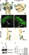
Conditional overexpression of β-catenin in ureteric cells results in severe renal defects. (A, B) Whole mount X-gal staining in E12.5 urogenital ridges resected from WT (A) and β-catGOF-UB mice (B) containing a ROSA-26 reporter allele. LacZ staining in Wt and β-catGOF-UB in E12.5 urogential ridges demonstrated Hoxb.7-Cre mediated excision in Wolffian duct (WD), mesonephric tubules (MT), and ureteric bud (UB) tissue. (C, D) β-catenin immunofluoresence in E12.5 WT and β-catGOF-UB kidney tissue. (C) In WT kidney β-catenin is localized in the UB plasma membrane consistent with a role in adherens junctions (white arrow). β-Catenin expression was not observed in the cytoplasm or nucleus in WT UBs. (D) In β-catGOF-UB mutant mice β-catenin localized to the ureteric cell plasma membrane as well as the cytoplasm and nucleus (white arrow). (E, F) Whole mount X-gal staining in E11.5 urogenital ridges isolated from WT TCF reporter mice and β-catGOF-UB;TCF mice demonstrates activity in the WD and UB. The X-gal staining in β-catGOF-UB;TCF mice is noticeably more intense when compared with WT littermates, thus indicating increased transcriptional activity. (G, H) Western analysis and corresponding densitometry for β-catenin and GAPDH in WT and β-catGOF-UB mutant kidneys.
Figure 2.
Figure 2.
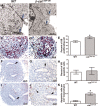
Arrested branching morphogenesis in Hoxb.7;β-catΔ3/+ mutant mice. (A, B) Urinary systems from P0 WT and β-catGOF-UB mice, demonstrating severe renal malformations in β-catGOF-UB mice (white arrows). (C, D) H&E stained kidney sections from P0 WT and β-catGOF-UB mice, demonstrating a lack of cortical medullary patterning, cystic tubules (C), and a paucity of nephrogenic structures. (E through H) H&E stained cross sections of mouse embryos at E11.5 and E12.5 demonstrates a paucity of ureteric bud (UB) tissue and nephrogenic elements as early as E12.5 in β-catGOF-UB mice. White arrow, MM; black arrow, UB tissue. (I, J) GFP fluorescence demonstrating arrested branching morphogenesis in β-catGOF-UB mice. MM, metanephric mesenchyme.
Figure 3.
Figure 3.
β-catenin stabilization modulates cellular events during ureteric branching. (A, B) Analysis of adherens junctions (white arrows) by transmission electron microscopy demonstrates the presence of junctional complexes in β-catGOF-UB tissue indistinguishable from WT. (C through E) Qualitative and quantitative analysis of cell proliferation in E12.5 WT and β-catGOF-UB kidney tissue using an in situ BrdU incorporation assay. Quantitative analysis of BrdU incorporation (red color) demonstrated a 1.24-fold increase (% BrdU-positive ureteric cells, WT [43.2%] versus β-catGOF-UB [53.7%], *P = 0.03) in β-catGOF-UB ureteric cell proliferation when compared with WT. (F through K) Qualitative and quantitative analysis of ureteric bud and mesenchymal apoptosis by TUNEL analysis. TUNEL-positive cells (brown color, arrowhead) are rarely detected in the ureteric bud (black arrow) in WT (F, G) and β-catGOF-UB (I, J) mutant mice (% TUNEL-positive ureteric cells, WT [0.33 ± 0.19%] versus β-catGOF-UB [0.40 ± 0.16%], P = 0.78). Apoptosis in the metanephric mesenchyme was increased 4.9-fold (number TUNEL-positive cells per mm2, 0.1 ± 0.02 versus 0.55 ± 0.12, *P = 0.015) in β-catGOF-UB mice when compared with that in WT (J, K).
Figure 4.
Figure 4.
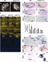
β-catenin stabilization in ureteric cells increases Tgfβ2 and DKK1 expression. (A, B) Ureteric bud–specific (UB-specific) green fluorescent protein fluorescence to identify the UB branching pattern in WT and β-catGOF-UB mice. Ureteric tissue is marked with a white arrow. WT and β-catGOF-UB mice were pooled to generate biologic triplicates. (C) Heat-map representation of differentially expressed genes between WT and β-catGOF-UB demonstrates a low level of variability of differentially expressed transcripts among sample replicates. Each lane represents individual biologic replicates normalized to each of three WT samples. (D through I) In situ hybridization for Gdnf, Ret, and Wnt11 in E12.5 WT and β-catGOF-UB kidneys. Gdnf was expressed in a mosaic pattern around the ureteric tips in β-catGOF-UB mice and Ret expression was limited to the tips of the UB tips in WT and β-catGOF-UB (black arrows). (H, I) In contrast to WT, Wnt11 was nearly absent in β-catGOF-UB ureteric tips (black arrows). (J) Validation of microarray data by quantitative real-time PCR confirmed a significant upregulation of Tgfβ2 (WT versus mutant: 0.013 ± 0.009 versus 0.067 ± 0.35, P = 0.03) and Dkk1 (0.9 ± 0.05 versus 1.41 ± 0.16. P = 0.038). (K through P) Spatial localization of the candidate genes. (K, L) Tgfβ2 mRNA is upregulated in E12.5 UB cells in β-catGOF-UB mutants. (M, N) Immunohistochemistry demonstrates a marked increase in Tgfβ2 protein expression in the metanephric mesenchyme (MM) in β-catGOF-UB mutants. (O,P) Dkk1 is markedly upregulated in E12.5 UB cells (UB) in β-catGOF-UB mutants. No changes in Tgfβ2 and Dkk1 mRNA were observed in MM cells.
Figure 5.
Figure 5.
Expanded and ectopic metanephric mesenchyme (MM) cell induction in β-catGOF-UB mutants. (A, B, E, F, I, J) Analysis of TGFβ2-treated E12.5 kidney explants and (C, D, G, H, K, L) kidney tissue isolated from E12.5 WT and β-catGOF-UB mice. (A through D) H&E stained kidney tissue in untreated and TGFβ2-treated kidney explants (A, B) and WT and β-catGOF-UB mutants (C, D) (black arrows). TGFβ2-treated (B) and β-catGOF-UB kidneys (D) demonstrate an expansion of mesenchymal aggregates around the tips of the UB (black arrows). (E through H) Dual-label immunofluorescence imaging of PAX2 and NCAM demonstrating expanded and ectopic NCAM and PAX2 expression in TGFβ2-treated kidney explants (E, F) and β-catGOF-UB mice (white arrow) (G, H). (I through L) Expression of CITED1, a nephrogenic lineage–specific marker, confirms an expansion of induced mesenchyme in TGFβ2-treated kidney explants (white arrow) (I, J). (K, L) CITED1 and NCAM co-immunofluorescence in WT and β-catGOF-UB mice confirms an expanded domain of cells expressing CITED1 adjacent to the UB (white arrows).
Figure 6.
Figure 6.
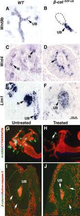
Disruption in nephrogenesis is Dkk1-dependent. (A through F) Analysis of genes necessary for nephrogenesis. Wnt9b localizes to the ureteric bud (UB) in E12.5 β-catGOF-UB mice in a pattern similar to WT (A, B). In contrast to WT, Wnt4 is ectopically expressed in the induced mesenchymal aggregates surrounding the ureteric tips in β-catGOF-UB mice (black arrow) (C, D). Lim1 mRNA is expressed in UB cells in both WT and β-catGOF-UB mutants. In contrast to WT, Lim1 mRNA expression is practically absent from the metanephric mesenchyme. (E, F). Resected E11.5 kidney explants were treated with DKK1 for 96 hours. CYTOKERATIN (red) demonstrates a similar ureteric branch pattern in untreated and DKK1-treated samples. In contrast, a noticeable decrease in brush boarder–positive structures (proximal tubules, green, white arrows) is observed in DKK1-treated samples (G, H). (I, J) Analysis of activated caspase-3 expression (apoptotic marker) in DKK1-treated explants. Activated caspase-3 expression is increased within metanephric mesenchyme in DKK1-treated kidney explants. n, nephrogenic structure.
Figure 7.
Figure 7.
Tamoxifen-induced overexpression of β-catenin in E14.5 embryos results in renal dysplasia. (A, D) H&E stained sections from resected E16.5 kidneys from WT and CreERT;β-catΔ3/+ mice. CreERT;β-catΔ3/+ mice demonstrate slightly smaller kidneys with an otherwise normal kidney morphology. In contrast to WT kidneys, CreERT;β-catΔ3/+ kidneys demonstrate an absence of a nephrogenic zone (black asterisk), a paucity of nephrogenic intermediate structures, and a deficiency developing and maturing glomeruli (D). Deep cortical glomeruli are observed in both WT and CreERT;β-catΔ3/+ mutant mice (black arrows). (E, F) CITED1-CYTOKERATIN dual immunofluorescence demonstrates ectopic CITED1 expression in the mesenchyme and tubule structures in E16.5 CreERT;β-catΔ3/+ mice (white arrows). In contrast, CYTOKERATIN was not observed in cortical tubules or medullary tubules in kidneys from CreERT;β-catΔ3/+ mice (white arrowhead). (G, H) PAX2 and NCAM co-immunofluorescence. PAX2 is expressed in renal tubules (T), and condensing mesenchyme (CM) around the tips of the UB in WT (white arrow, CM). Similar to WT mice, PAX2 is expressed in renal tubules, but is virtually absent in the cortical mesenchyme in CreERT;β-catΔ3/+ mice (white asterisk). A similar pattern of NCAM expression was observed in WT and CreERT;β-catΔ3/+. (I, J) In situ hybridization for Tgfβ2 demonstrates an increase in mRNA expression in tubules localized in the cortex and medullary regions in CreERT;β-catΔ3/+ kidneys when compared with WT (black arrows and highlighted in inset boxes). (K, L) Immunohistochemistry demonstrates increased Tgfβ2 protein expression in glomeruli and tubules in CreERT;β-catΔ3/+mice. Tgfβ2 protein expression was also observed in the condensing mesenchyme surrounding the UB (highlighted in inset boxes). (M, N) In situ hybridization for Dkk1 demonstrates an obvious increase in mRNA expression in cortex and medullary tubules (black arrow) in CreERT;β-catΔ3/+ kidneys when compared with WT (black asterisk). DG, developing glomeruli; G, glomeruli; CM, condensing mesenchyme; T, tubules; NZ, nephrogenic zone.
Figure 8.
Figure 8.
Effects of β-catenin overexpression during kidney embryogenesis. Overexpression of β-catenin in ureteric cells during embryogenesis increases Tgfβ2 and Dkk1. Tgfβ2 acts in an autocrine manner to inhibit ureteric branching. In addition, Tgfβ2 acts in a paracrine manner to induce the formation of an ectopic population of mesenchyme cells committed a nephrogenic fate. Progression of nephrogenesis within this ectopic population of cells, as well as those induced by adjacent ureteric cells, is inhibited by the simultaneous paracrine actions of DKK1. CM, condensing mesenchyme; MM, metanephric mesenchyme; RV, renal vesicle; WD, Wolffian duct; UB, ureteric bud; UBM, ureteric branching morphogenesis.

Comment in

Similar articles

Cited by

References

    1. N'Guessen G, Stephens FD, Pick J: Congenital superior ectopic (thoracic) kidney. Urology 24: 219–228, 1984 - PubMed
    1. Saxen L, Sariola H: Early organogenesis of the kidney. Pediatr Nephrol 1: 385–392, 1987 - PubMed
    1. Piscione TD, Rosenblum ND: The malformed kidney: Disruption of glomerular and tubular development. Clin Genet 56: 341–356, 1999 - PubMed
    1. Majumdar A, Vainio S, Kispert A, McMahon J, McMahon AP: Wnt11 and Ret/Gdnf pathways cooperate in regulating ureteric branching during metanephric kidney development. Development 130: 3175–3185, 2003 - PubMed
    1. Carroll TJ, Park JS, Hayashi S, Majumdar A, McMahon AP: Wnt9b plays a central role in the regulation of mesenchymal to epithelial transitions underlying organogenesis of the mammalian urogenital system. Dev Cell 9: 283–292, 2005 - PubMed

Publication types

MeSH terms