. 2011 Apr 8;332(6026):247-51.
doi: 10.1126/science.1201678. Epub 2011 Mar 24.

AMP-activated protein kinase regulates neuronal polarization by interfering with PI 3-kinase localization

Affiliations

AMP-activated protein kinase regulates neuronal polarization by interfering with PI 3-kinase localization

Stephen Amato et al. Science. .

Abstract

Axon-dendrite polarization is crucial for neural network wiring and information processing in the brain. Polarization begins with the transformation of a single neurite into an axon and its subsequent rapid extension, which requires coordination of cellular energy status to allow for transport of building materials to support axon growth. We found that activation of the energy-sensing adenosine 5'-monophosphate (AMP)-activated protein kinase (AMPK) pathway suppressed axon initiation and neuronal polarization. Phosphorylation of the kinesin light chain of the Kif5 motor protein by AMPK disrupted the association of the motor with phosphatidylinositol 3-kinase (PI3K), preventing PI3K targeting to the axonal tip and inhibiting polarization and axon growth.


Figures

Fig. 1
Fig. 1
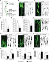
AMPK activation suppresses neuronal polarization in cultured hippocampal neurons. (A) Time course for AMPK activation (pAMPK) by AICAR (2 mM) in day 1 in vitro (DIV1) hippocampal neurons. (B) Incubation of DIV1 neurons with AICAR and metformin (200 µM) on AMPK activation. (C) Neurons were pretreated with compound C (CC) (20 µM) for 1 hour followed by 30-min incubation with AICAR. (D) Illustration of AICAR treatment paradigm with corresponding polarization stages. (E to G) Effect of AMPK activation by AICAR and metformin on neuronal polarization. (H to J) Measurement of nonaxonal neurite length, axon length, and the total growth. (K and L) Expression of wild-type (WT) or kinase-dead (KD) AMPK on neuronal polarity. Cell numbers of statistical analysis are indicated at the top of bar graphs. *P < 0.05, two-population Student’s t test [(F) and (H) to (J)], or analysis of variance (ANOVA) with Tukey post-test [(G) and (L)]. Scale bar, 20 µm.
Fig. 2
Fig. 2
AMPK activation disrupts tip localization of active Akt. (A and B) AICAR treatment on PI3K-mediated Akt phosphorylation in DIV1 neurons. (C) Illustration of AICAR treatment paradigm. (D)Hippocampal neurons treated with AICAR and LY294002 (LY), respectively, or together for 15 hours, immunostained for phospho-Akt (Ser473) and neuronal marker Tuj1. Scale bar, 20 µm. (E) Quantification of tip fluorescence intensity normalized to shaft intensity. (F) Normalization of tip pAkt intensity to tip area indicated by EGFP. (G) EGFP intensity at axonal tip. (H) Quantification of pAkt soma intensity. (I) Plot profile of p-Akt signals from the soma to neurite tips. Dashed line indicates background intensity. (J) Live imaging of PH-EGFP in stage 3 neurons. *P < 0.05, ANOVA with Tukey post-test [(E) and (H)], or two-population Student’s t test [(F) and (G)]. Scale bar, 10 µm.
Fig. 3
Fig. 3
Association of KLC2 and p85 is required for PI3K tip enrichment and neuronal polarity. (A) In vitro phosphorylation of KLC2 by AMPK. Purified GST-KLC2 was incubated with wild-type (WT) or kinase dead (KD) AMPK catalytic α1 subunit in the presence of P32-labeled ATP. Positive radioactivity was observed in KLC2 and AMPK α1 subunit, indicating phosphorylation of KLC2 and autophosphorylation of AMPK. GST (glutathione S-transferase) was used as a control. (B) Immunoprecipitation of GFP-p85 in human embryonic kidney cells expressing GFP-p85 together with wild-type or mutant Myc-KLC2 (DM). (C to E) Immunostaining of p85 in stage 3 hippocampal neurons transfected with EGFP alone or together with either the WT or DM KLC2. (F and G) AICAR treatment on neurons expressing EGFP together with WT or DM KLC2. *P < 0.05, ANOVA with Tukey post-test. Scale bar, 20 µm.
Fig. 4
Fig. 4
AMPK activation disrupts neuronal polarization in organotypic slice culture. (A) Illustration of experimental design and stages of neuronal polarization. (B to F) Cortical brain slices prepared from E14 mice were infected with CAG-GFP retrovirus to indicate newborn neurons and treated with AICAR (2 mM, 72 hours). (G to M) The embryonic brain (E15.5) was in utero–electroporated with EGFP with or without AMPK-KD, and brain slices were prepared for AICAR incubation (48 hours). Arrowheads and stars indicate axonal segment and the soma, respectively. *P < 0.05, two-population Student’s t test [(D) to (F)], or ANOVA with Tukey post-test [(K) to (M)]. Scale bar, 10 µm.

References

    1. Jareb M, Banker G. J. Neurosci. 1997;17:8955. - PMC - PubMed
    1. Hardie DG, Hawley SA, Scott JW. J. Physiol. 2006;574:7. - PMC - PubMed
    1. Dasgupta B, Milbrandt J. Dev. Cell. 2009;16:256. - PMC - PubMed
    1. Sabina RL, Patterson D, Holmes EW. J. Biol. Chem. 1985;260:6107. - PubMed
    1. Dotti CG, Sullivan CA, Banker GA. J. Neurosci. 1988;8:1454. - PMC - PubMed

Publication types

MeSH terms