. 2011 Jul;18(7):457-66.
doi: 10.1038/cgt.2011.10. Epub 2011 Mar 25.

In tumors Salmonella migrate away from vasculature toward the transition zone and induce apoptosis

Affiliations

In tumors Salmonella migrate away from vasculature toward the transition zone and induce apoptosis

S Ganai et al. Cancer Gene Ther. 2011 Jul.

Abstract

Motile bacteria can overcome diffusion resistances that substantially reduce the efficacy of standard cancer therapies. Many reports have also recently described the ability of Salmonella to deliver therapeutic molecules to tumors. Despite this potential, little is known about the spatiotemporal dynamics of bacterial accumulation in solid tumors. Ultimately this timing will affect how these microbes are used therapeutically. To determine how bacteria localize, we intravenously injected Salmonella typhimurium into BALB/c mice with 4T1 mammary carcinoma and measured the average bacterial content as a function of time. Immunohistochemistry was used to measure the extent of apoptosis, the average distance of bacteria from tumor vasculature and the location of bacteria in four different regions: the core, transition, body and edge. Bacteria accumulation was also measured in pulmonary and hepatic metastases. The doubling time of bacterial colonies in tumors was measured to be 16.8 h, and colonization was determined to delay tumor growth by 48 h. From 12 and 48 h after injection, the average distance between bacterial colonies and functional vasculature significantly increased from 130 to 310 μm. After 48 h, bacteria migrated away from the tumor edge toward the central core and induced apoptosis. After 96 h, bacteria began to marginate to the tumor transition zone. All observed metastases contained Salmonella and the extent of bacterial colocalization with metastatic tissue was 44% compared with 0.5% with normal liver parenchyma. These results demonstrate that Salmonella can penetrate tumor tissue and can selectively target metastases, two critical characteristics of a targeted cancer therapeutic.


Figures
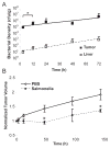
Figure 1
Figure 1. Salmonella proliferated exponentially in 4T1 mammary tumors
A, bacterial density(cfu/g) in tumors and liver samples harvested after systemic injection of S. typhimurium. Density was greater at 12 hours compared to 3 hours (*, p<0.05). Exponential growth was observed in both tissues. B, Tumor volume was significantly less in tumors treated with S. typhimurium at 48 and 144 hours compared to saline controls (*, p<0.05). Tumor volumes were normalized to initial volume.
Figure 2
Figure 2. Salmonella accumulated in distinct tumor regions over time
A, Composite images of 4T1 Tumors, 12 hours after systemic bacterial injection. Serial 4μm sections were dual label stained using a combination of Salmonella immunohistochemistry and biotinylated tomato lectin (left) to visualize bacteria (red) and perfused vasculature (brown) or stained using cleaved caspase-3 immunohistochemistry (middle) to visualize regions of apoptosis (brown). Scale bars are 1mm. Based on histological staining, four regions with distinct microenvironments were observed in each tumor: edge (within 1mm of tumor periphery), body, transition, or core. B, Salmonella accumulated as colonies in the edge (left) and body (middle left) tumor regions. Bacteria were seen dispersed throughout the extracellular space, shown here in the body region (middle right). Arrows indicate colony edges (left, middle left) and individual bacteria in the extracellular space (middle right). Salmonella were observed both extracellularly and intracellularly (white arrows, right). The black arrow highlights a single extracellular bacterium (right). Scale bars are 50μm (left, middle left), 25μm (middle right), and 10μm (right). C, Dynamics of bacterial accumulation were determined by measuring pixel density per image and normalized to the edge microenvironment at 12 hours. At 12 and 48 hours, there was significantly greater accumulation within the edge than all other regions (*, p<0.05; †, p<0.01; except the core at 48 hrs). At 96 hours, there was greater accumulation within the tumor transition zone compared to all other regions (‡, p<0.01). D, At late time points Salmonella accumulated as stratified bands in the tumor transition zone (between arrows, left and highlighted, right). Arrows indicate boundary of the transition zone. Sparse accumulation in the necrotic core is present in D-left. Scale bars are 1mm (left) and 100μm (right).
Figure 3
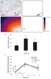
Figure 3. Systemic bacterial injection induced apoptosis in 4T1 tumors
A, Composite images of equatorial tumor sections stained for cleaved caspase-3. Sections were oriented in a coronal plane along craniocaudal and medial/lateral axes, and imaged using the blue channel filter. B, Individual images in each tumor composite (as in A) were scored as apoptotic, necrotic or viable based on identification of pyknosis, nuclear condensation or cleaved caspase-3. At 48 hours, the extent of apoptosis significantly increased and the extent of viable tissue decreased compared to PBS controls (*, p<0.05). No differences were seen in the relative proportion of tumor necrosis. C, Significantly more apoptotic tissue was observed 48 hours after injection in the transition and core regions compared to PBS controls (*, p<0.05). D, For all tumors, greater colocalization of bacteria with apoptosis was seen in the transition zone compared to the edge (*, p<0.05).
Figure 4
Figure 4. Bacterial accumulation migrated away from vessels with time
A, The location of Salmonella colonies (white arrow, red stain) and perfused blood vessels (black arrow, brown stain) were identified using dual label immunohistochemistry (upper left). The location of vessels was captured from the blue component of the image (upper right) and used to generate a Euclidian distance map (lower left). Merged distance maps identified the location of bacterial colonies relative to the nearest functional vessel (lower right). The mean distance between the colonies and perfused vasculature in this example image is 185.9 ± 67.6 (SD) μm. The scale bar is 25μm. B, The mean distance between bacteria and vasculature increased for 48hrs after injection. The distances at 12 and 48 hrs were greater than zero (*, p<0.05) and increased between 12 and 48hrs (†, p<0.0001). Thirty images were examined per tumor. C, Maximum distance of bacteria from vasculature as a function of time, grouped by region. At 12 hours, the maximum distance was greater in the core compared to the edge (*, p<0.05). The maximum distance in the edge increased from 12 to 48 hours (*, p<0.01). For clarity, error bars are only displayed for the edge and core regions.
Figure 5
Figure 5. Salmonella targets breast cancer metastases
Contiguous hepatic (A, B) and pulmonary (C, D) sections were stained with hematoxylin and eosin (A, C) and Salmonella immunohistochemistry (brown in B, D). A–B, Salmonella accumulated in hepatic metastases with greater specificity than normal liver parenchyma. Bacterial colonies (boundary indicated with dark arrows in B) colocalized with metastatic tissue (boundary indicated with white arrows in A). Scale bars are 250μm. C–D, Salmonella (e.g. white arrow) accumulated in small pulmonary micro-metastases. The micro-metastasis encased a vascular channel containing an inflammatory infiltrate (black arrow). Scale bars are 50μm.
Figure 6
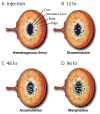
Figure 6. Model of Salmonella accumulation within tumor microenvironments
A) Bacteria first access the tumor via hematogenous entry. B) During the first 12 hours bacteria disseminate throughout all microenvironment regions. C) For first 48 hours bacteria migrate and proliferate, resulting to accumulation within all regions. D) By 96 hours, bacteria marginate to the tumor transition zone and density decreases in other regions.

References

    1. Sporn MB. The war on cancer. Lancet. 1996;347(9012):1377–81. - PubMed
    1. Cairns R, Papandreou I, Denko N. Overcoming physiologic barriers to cancer treatment by molecularly targeting the tumor microenvironment. Mol Cancer Res. 2006;4(2):61–70. - PubMed
    1. Jain RK, Forbes NS. Can engineered bacteria help control cancer? Proc Natl Acad Sci U S A. 2001;98(26):14748–50. - PMC - PubMed
    1. Minchinton AI, Tannock IF. Drug penetration in solid tumours. Nat Rev Cancer. 2006;6(8):583–92. - PubMed
    1. Albini A, Sporn MB. The tumour microenvironment as a target for chemoprevention. Nat Rev Cancer. 2007;7(2):139–47. - PubMed

Publication types