. 2011:2011:765923.
doi: 10.1155/2011/765923. Epub 2011 Feb 22.

Blood-brain barrier breakdown following traumatic brain injury: a possible role in posttraumatic epilepsy

Affiliations

Blood-brain barrier breakdown following traumatic brain injury: a possible role in posttraumatic epilepsy

Oren Tomkins et al. Cardiovasc Psychiatry Neurol. 2011.

Abstract

Recent animal experiments indicate a critical role for opening of the blood-brain barrier (BBB) in the pathogenesis of post-traumatic epilepsy (PTE). This study aimed to investigate the frequency, extent, and functional correlates of BBB disruption in epileptic patients following mild traumatic brain injury (TBI). Thirty-seven TBI patients were included in this study, 19 of whom suffered from PTE. All underwent electroencephalographic (EEG) recordings and brain magnetic resonance imaging (bMRI). bMRIs were evaluated for BBB disruption using novel quantitative techniques. Cortical dysfunction was localized using standardized low-resolution brain electromagnetic tomography (sLORETA). TBI patients displayed significant EEG slowing compared to controls with no significant differences between PTE and nonepileptic patients. BBB disruption was found in 82.4% of PTE compared to 25% of non-epileptic patients (P = .001) and could be observed even years following the trauma. The volume of cerebral cortex with BBB disruption was significantly larger in PTE patients (P = .001). Slow wave EEG activity was localized to the same region of BBB disruption in 70% of patients and correlated to the volume of BBB disrupted cortex. We finally present a patient suffering from early cortical dysfunction and BBB breakdown with a gradual and parallel resolution of both pathologies. Our findings demonstrate that BBB pathology is frequently found following mild TBI. Lasting BBB breakdown is found with increased frequency and extent in PTE patients. Based on recent animal studies and the colocalization found between the region of disrupted BBB and abnormal EEG activity, we suggest a role for a vascular lesion in the pathogenesis of PTE.


Figures

Figure 1
Figure 1
Imaging modalities allow for BBB permeability evaluation and quantification. (a) Control subject: no BBB disruption is observed within the brain parenchyma (areas of significant differences are found in blood vessels, sinuses and the choroid plexus). Increased BBB permeability (K trans) and extravascular volume (v e) are also localized to those same structures. (b) A 28-year-old PTE patient 10 days following TBI. A region of increased BBB permeability is detected over the left parietal lobe in both methods (arrows).
Figure 2
Figure 2
Abnormal qEEG in TBI patients. (a) Average normalized power spectrum representation of all TBI patients and controls that underwent qEEG. Note the significant increase in delta power and the decrease in the alpha band in TBI patients compared to controls (inset). (b) Power spectrum averages of patient subgroups according to the occurrence of seizures. While both the PTE and the non-epileptic group had elevated delta power compared to controls, only PTE patients had a significant reduction of alpha and increase of theta power. *= P < .05.
Figure 3
Figure 3
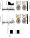
The extent of qEEG slowing in PTE patients correlates with BBB disruption. (a) An enlarged region of normalized sLORETA values for the delta band in voxels number 3900–4100 from healthy controls. The entire normalized sLORETA for the delta band is shown in the inset, as are 2 examples of signal localization to the frontal midline region. (b) The PTE population displayed marked variability among the same voxels, and maximal signal localization was varied according to the site of injury. (c) The volume of cortex with abnormal cortical activity according to sLORETA between patients with the bottom (black) and top (white) half volume of BBB disruption or cortical lesion. Note that patients with a larger volume of BBB disruption also had a significantly larger volume of dysfunctional cortex. *= P < .05.
Figure 4
Figure 4
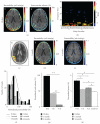
Abnormal EEG slowing localizes to region of BBB disruption in a 15-year-old PTE patient one month following mild TBI (see text for details). (a) BBB evaluation 10 days after the event revealed a focal area of increased BBB permeability (left, arrow, Brodmann area 40), surrounded by an increase of extravascular volume (right, arrow). (b) Representation of the EEG recording with the x-axis representing the 23 different electrodes, the y-axis the frequencies at 0.5 Hz intervals, and the colour coding the number of standard deviations from the average control EEG. Note the increased power in the delta range over the right temporoparietal electrodes (arrows). (c) sLORETA localized the delta activity to the right parietal region (Brodmann area 40). Repeated MRI scans 1 (d) and 4 months (e) following the trauma revealed a resolution of the BBB lesion. (f) Histogram representation of the permeability values surrounding the cortical lesion (normalized to the average value of the contralateral hemisphere). (g) Four months after the trauma there is a significant decrease in the permeability values, as well as in the extravascular volume. (h) Quantification of the average delta power shows a significant reduction in delta wave activity as time progresses, though remaining significantly increased compared to controls even 4 months after the event and resolution of the BBB lesion. * = P < .05, ** = P < .0001.

References

    1. Annegers JF, Grabow JD, Groover RV, Laws ER, Jr., Elveback LR, Kurland LT. Seizures after head trauma: a population study. Neurology. 1980;30(7):683–689. - PubMed
    1. Annegers JF, Hauser WA, Coan SP, Rocca WA. A population-based study of seizures after traumatic brain injuries. The New England Journal of Medicine. 1998;338(1):20–24. - PubMed
    1. Desai BT, Whitman S, Coonley Hoganson R, Coleman TE, Gabriel G, Dell J. Seizures and civilian head injuries. Epilepsia. 1983;24(3):289–296. - PubMed
    1. Caveness WF, Meirowsky AM, Rish BL, et al. The nature of posttraumatic epilepsy. Journal of Neurosurgery. 1979;50(5):545–553. - PubMed
    1. Asikainen I, Kaste M, Sarna S. Early and late posttraumatic seizures in traumatic brain injury rehabilitation patients: brain injury factors causing late seizures and influence of seizures on long-term outcome. Epilepsia. 1999;40(5):584–589. - PubMed

LinkOut - more resources