

Effects of anti-VEGF on predicted antibody biodistribution: roles of vascular volume, interstitial volume, and blood flow
- PMID: 21436893
- PMCID: PMC3060062
- DOI: 10.1371/journal.pone.0017874
Effects of anti-VEGF on predicted antibody biodistribution: roles of vascular volume, interstitial volume, and blood flow
Abstract
Background: The identification of clinically meaningful and predictive models of disposition kinetics for cancer therapeutics is an ongoing pursuit in drug development. In particular, the growing interest in preclinical evaluation of anti-angiogenic agents alone or in combination with other drugs requires a complete understanding of the associated physiological consequences.
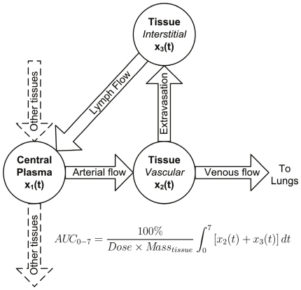
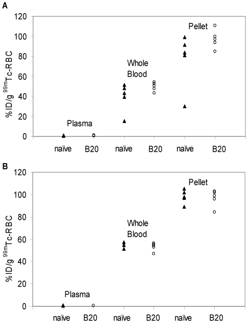
Methodology/principal findings: Technescan™ PYP™, a clinically utilized radiopharmaceutical, was used to measure tissue vascular volumes in beige nude mice that were naïve or administered a single intravenous bolus dose of a murine anti-vascular endothelial growth factor (anti-VEGF) antibody (10 mg/kg) 24 h prior to assay. Anti-VEGF had no significant effect (p>0.05) on the fractional vascular volumes of any tissues studied; these findings were further supported by single photon emission computed tomographic imaging. In addition, apart from a borderline significant increase (p = 0.048) in mean hepatic blood flow, no significant anti-VEGF-induced differences were observed (p>0.05) in two additional physiological parameters, interstitial fluid volume and the organ blood flow rate, measured using indium-111-pentetate and rubidium-86 chloride, respectively. Areas under the concentration-time curves generated by a physiologically-based pharmacokinetic model changed substantially (>25%) in several tissues when model parameters describing compartmental volumes and blood flow rates were switched from literature to our experimentally derived values. However, negligible changes in predicted tissue exposure were observed when comparing simulations based on parameters measured in naïve versus anti-VEGF-administered mice.
Conclusions/significance: These observations may foster an enhanced understanding of anti-VEGF effects in murine tissues and, in particular, may be useful in modeling antibody uptake alone or in combination with anti-VEGF.
Conflict of interest statement
Figures


Similar articles
-
Development and evaluation of a novel method for preclinical measurement of tissue vascular volume.Mol Pharm. 2010 Oct 4;7(5):1848-57. doi: 10.1021/mp100183k. Epub 2010 Aug 12. Mol Pharm. 2010. PMID: 20704296
-
Effects of anti-VEGF on pharmacokinetics, biodistribution, and tumor penetration of trastuzumab in a preclinical breast cancer model.Mol Cancer Ther. 2012 Mar;11(3):752-62. doi: 10.1158/1535-7163.MCT-11-0742-T. Epub 2012 Jan 5. Mol Cancer Ther. 2012. PMID: 22222630
-
Antibody neutralization of vascular endothelial growth factor inhibits wound granulation tissue formation.J Surg Res. 2001 Apr;96(2):173-82. doi: 10.1006/jsre.2001.6089. J Surg Res. 2001. PMID: 11266270
-
Vascular endothelial growth factor (VEGF) as a target of bevacizumab in cancer: from the biology to the clinic.Curr Med Chem. 2006;13(16):1845-57. doi: 10.2174/092986706777585059. Curr Med Chem. 2006. PMID: 16842197 Review.
-
Improving delivery of antineoplastic agents with anti-vascular endothelial growth factor therapy.Cancer. 2005 Apr 15;103(8):1561-70. doi: 10.1002/cncr.20942. Cancer. 2005. PMID: 15754332 Review.
Cited by
-
Tissue distribution studies of protein therapeutics using molecular probes: molecular imaging.AAPS J. 2012 Sep;14(3):389-99. doi: 10.1208/s12248-012-9348-3. Epub 2012 Mar 31. AAPS J. 2012. PMID: 22467336 Free PMC article. Review.
-
Pre-clinical Evaluation of a Cyanine-Based SPECT Probe for Multimodal Tumor Necrosis Imaging.Mol Imaging Biol. 2016 Dec;18(6):905-915. doi: 10.1007/s11307-016-0972-7. Mol Imaging Biol. 2016. PMID: 27277828 Free PMC article.
-
Tissue Physiology of Cynomolgus Monkeys: Cross-Species Comparison and Implications for Translational Pharmacology.AAPS J. 2018 Oct 8;20(6):107. doi: 10.1208/s12248-018-0264-z. AAPS J. 2018. PMID: 30298434
-
Preclinical pharmacokinetics, pharmacodynamics, tissue distribution, and tumor penetration of anti-PD-L1 monoclonal antibody, an immune checkpoint inhibitor.MAbs. 2016;8(3):593-603. doi: 10.1080/19420862.2015.1136043. Epub 2016 Feb 26. MAbs. 2016. PMID: 26918260 Free PMC article.
-
Relaxin improves multiple markers of wound healing and ameliorates the disturbed healing pattern of genetically diabetic mice.Clin Sci (Lond). 2013 Dec;125(12):575-85. doi: 10.1042/CS20130105. Clin Sci (Lond). 2013. Retraction in: Clin Sci (Lond). 2024 Nov 20;138(22):1503. doi: 10.1042/CS-2013-0105_RET. PMID: 23742173 Free PMC article. Retracted.
References
-
- Boswell CA, Deng R, Lin K, Putnam WS, Lei C, et al. In vitro-in vivo correlations of pharmacokinetics, pharmacodynamics and metabolism for antibody therapeutics. In: Mrsny RJ, Daugherty A, editors. Proteins and Peptides: Pharmacokinetic, Pharmacodynamic, and Metabolic Outcomes. New York, NY: Informa HealthCare; 2009.
-
- Boswell CA, Ferl GZ, Mundo EE, Schweiger MG, Marik J, et al. Development and Evaluation of a Novel Method for Preclinical Measurement of Tissue Vascular Volume. Mol Pharm. 2010;7:1848–1857. - PubMed
-
- Brown RP, Delp MD, Lindstedt SL, Rhomberg LR, Beliles RP. Physiological parameter values for physiologically based pharmacokinetic models. Toxicol Ind Health. 1997;13:407–484. - PubMed
-
- Davies B, Morris T. Physiological parameters in laboratory animals and humans. Pharm Res. 1993;10:1093–1095. - PubMed
-
- Baxter LT, Zhu H, Mackensen DG, Jain RK. Physiologically based pharmacokinetic model for specific and nonspecific monoclonal antibodies and fragments in normal tissues and human tumor xenografts in nude mice. Cancer Res. 1994;54:1517–1528. - PubMed
MeSH terms
Substances
LinkOut - more resources
Full Text Sources
Miscellaneous
