. 2010 Oct;5(5):645-661.
doi: 10.1586/eop.10.63.

Update on amniotic membrane transplantation

Affiliations

Update on amniotic membrane transplantation

Jingbo Liu et al. Expert Rev Ophthalmol. 2010 Oct.

Abstract

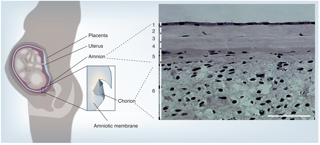
Cryopreserved amniotic membrane modulates adult wound healing by promoting epithelialization while suppressing stromal inflammation, angiogenesis and scarring. Such clinical efficacies of amniotic membrane transplantation have been reported in several hundred publications for a wide spectrum of ophthalmic indications. The success of the aforementioned therapeutic actions prompts investigators to use amniotic membrane as a surrogate niche to achieve ex vivo expansion of ocular surface epithelial progenitor cells. Further investigation into the molecular mechanism whereby amniotic membrane exerts its actions will undoubtedly reveal additional applications in the burgeoning field of regenerative medicine. This article will focus on recent advances in amniotic membrane transplantation and expand to cover its clinical uses beyond the ocular surface.


Figures

Figure 1
Figure 1. The amniotic membrane in the uterus and its histology
The placental membrane consists of the outer chorion (depicted in dark grey) and the inner amniotic membrane (depicted in light grey). Histologically, amniotic membrane is composed of (1) a monolayer of simple epithelium with a basement membrane, and an avascular stroma, which can further be subdivided into (2) compact, (3) fibroblast, (4) sponge and (5) reticular layers. Amniotic membrane is fused with (6) the cytotrophoblast layer of the chorion.
Figure 2
Figure 2. Publications released between 1995 and 2009 describing the use of amniotic membrane transplantation in ophthalmology
Figure 3
Figure 3. Prokera® and its application in ocular surface
(A) ProKera® is a dual-ring system that fastens a sheet of semitransparent cryopreserved amniotic membrane. It enhances the ease of patient care in many difficult corneal diseases. It is inserted under topical anesthesia into the upper fornix first, and then tucked under the lower lid. (B) A slit-lamp photograph depicts its appearance when inserted in the eye. (C) The status of epithelialization can be monitored by fluorescein staining and the intraocular pressure can be measured by Tonopen™ without having ProKera removed.
Figure 4
Figure 4. Amniotic membrane transplant ‘stagewise approach’ for nearly total limbal stem cell deficiency
(A) The first-stage surgery directed to conjunctivalized pannus marked by broken lines resulted in (B) a full recovery of the limbal and corneal surfaces 3 months after surgery. (B) The second-stage surgery was directed to the residual pannus from the remaining less-involved areas marked by broken lines. (C) As a result, the cornea recovered a stable and smooth epithelium without vascularization and much less cloudiness 21 months later. Reprinted with permission from [106].
Figure 5
Figure 5. The three surgical strategies for fornix reconstruction
(A) After symblepharon lysis and removal of subconjunctival scar tissue, amniotic membrane (pink) is used to cover the denuded scleral surface with fibrin glue or sutures up to the recessed conjunctiva (green) in mild symblepharon. (B) One anchoring suture (gray) per quadrant is used to secure the recessed conjunctival edge (green) to the skin with a bolster (gray circle) in moderate symblepharon. (C) Additional oral mucosa (orange) is used to extend the epithelial covering from the (green) residual conjunctiva in severe symblepharon. Reproduced with permission from [141].
Figure 6
Figure 6. Representative intraoperative (A & B) and postoperative (C & D) photographs of amniotic membrane (A & C) and pericardium (B & D) covering the glaucoma drainage shunt tube

References

    1. Tseng SCG, Espana EM, Kawakita T, et al. How does amniotic membrane work? Ocul. Surf. 2004;2(3):177–187. - PubMed
    1. Sippel KC, Ma JJK, Foster CS. Amniotic membrane surgery. Curr. Opin. Ophthalmol. 2001;12:269–281. - PubMed
    1. Tseng SCG. Amniotic membrane transplantation for ocular surface reconstruction. Bioscience Rep. 2002;21:481–489. - PubMed
    1. Dua HS, Gomes JA, King AJ, Maharajan VS. The amniotic membrane in ophthalmology. Surv. Ophthalmol. 2004;49(1):51–77. - PubMed
    1. Bouchard CS, John T. Amniotic membrane transplantation in the management of severe ocular surface disease: indications and outcomes. Ocul. Surf. 2004;2(3):201–211. - PubMed

LinkOut - more resources