Pancreatic serous cystadenoma with compression of the main pancreatic duct: an unusual entity
- PMID: 21436987
- PMCID: PMC3062951
- DOI: 10.1155/2011/574378
Pancreatic serous cystadenoma with compression of the main pancreatic duct: an unusual entity
Abstract
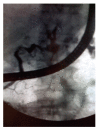
Serous cystadenoma is a common benign neoplasm that can be managed without surgery in asymptomatic patients provided that the diagnosis is certain. We describe a patient, whose pancreatic cyst exhibited a radiological appearance distinct from that of typical serous cystadenoma, resulting in diagnostic difficulties. CT and MRI showed a 10 cm-polycystic tumor with upstream dilatation of the main pancreatic duct (MPD), suggestive of intraductal papillary mucinous tumor (IPMT). Ultrasonographic aspect and EUS-guided fine-needle aspiration gave arguments for serous cystadenoma. ERCP showed a communication between cysts and the dilated MPD, compatible with IPMT. The patient underwent left pancreatectomy with splenectomy. Pathological examination concluded in a serous cystadenoma, with only a ductal obstruction causing proximal dilatation.
Figures


Similar articles
-
Macrocystic neoplasms of the pancreas: CT differentiation of serous oligocystic adenoma from mucinous cystadenoma and intraductal papillary mucinous tumor.AJR Am J Roentgenol. 2006 Nov;187(5):1192-8. doi: 10.2214/AJR.05.0337. AJR Am J Roentgenol. 2006. PMID: 17056905
-
Serous cystadenoma in communication with the pancreatic duct: an unusual radiologic and pathologic entity.J Clin Gastroenterol. 2010 Jul;44(6):e133-5. doi: 10.1097/MCG.0b013e3181d3458d. J Clin Gastroenterol. 2010. PMID: 20216080
-
[A case of serous cystadenoma of the pancreas communicating with the main pancreatic duct synchronously diagnosed with pancreatic ductal carcinoma].Nihon Shokakibyo Gakkai Zasshi. 2013 Mar;110(3):449-55. Nihon Shokakibyo Gakkai Zasshi. 2013. PMID: 23459540 Japanese.
-
Cystic and ductal tumors of the pancreas: diagnosis and management.J Visc Surg. 2013 Apr;150(2):69-84. doi: 10.1016/j.jviscsurg.2013.02.003. Epub 2013 Mar 19. J Visc Surg. 2013. PMID: 23518192 Review.
-
Mucin-secreting tumors of the pancreas.Gastrointest Endosc Clin N Am. 1995 Jan;5(1):237-58. Gastrointest Endosc Clin N Am. 1995. PMID: 7728346 Review.
Cited by
-
A disseminated variant of pancreatic serous cystadenoma causing obstructive jaundice, a very rare entity: a case report and review of the literature.BMC Res Notes. 2014 Oct 22;7:749. doi: 10.1186/1756-0500-7-749. BMC Res Notes. 2014. PMID: 25338636 Free PMC article. Review.
References
-
- Schulz HU, Kellner U, Kahl S, et al. A giant pancreatic serous microcystic adenoma with 20 years follow-up. Langenbeck’s Archives of Surgery. 2007;392(2):209–213. - PubMed
-
- Kim SY, Lee JM, Kim SH, et al. Macrocystic neoplasms of the pancreas: CT differentiation of serous oligocystic adenoma from mucinous cystadenoma and intraductal papillary mucinous tumor. American Journal of Roentgenology. 2006;187(5):1192–1198. - PubMed
-
- Inoue S, Yamaguchi K, Shimizu S, et al. Serous cystadenoma of the pancreas with atypical imaging features: a new variant of serous cystadenoma of the pancreas? Pancreas. 1998;16(1):102–105. - PubMed
-
- Colonna J, Plaza JA, Frankel WL, Yearsley M, Bloomston M, Marsh WL. Serous cystadenoma of the pancreas: clinical and pathological features in 33 patients. Pancreatology. 2008;8(2):135–141. - PubMed
-
- Choi JY, Kim MJ, Lee JY, et al. Typical and atypical manifestations of serous cystadenoma of the pancreas: imaging findings with pathologic correlation. American Journal of Roentgenology. 2009;193(1):136–142. - PubMed
LinkOut - more resources
Full Text Sources

