. 2011 Feb 23:2011:715341.
doi: 10.4061/2011/715341.

Amniotic-Fluid Stem Cells: Growth Dynamics and Differentiation Potential after a CD-117-Based Selection Procedure

Affiliations

Amniotic-Fluid Stem Cells: Growth Dynamics and Differentiation Potential after a CD-117-Based Selection Procedure

S Arnhold et al. Stem Cells Int. .

Abstract

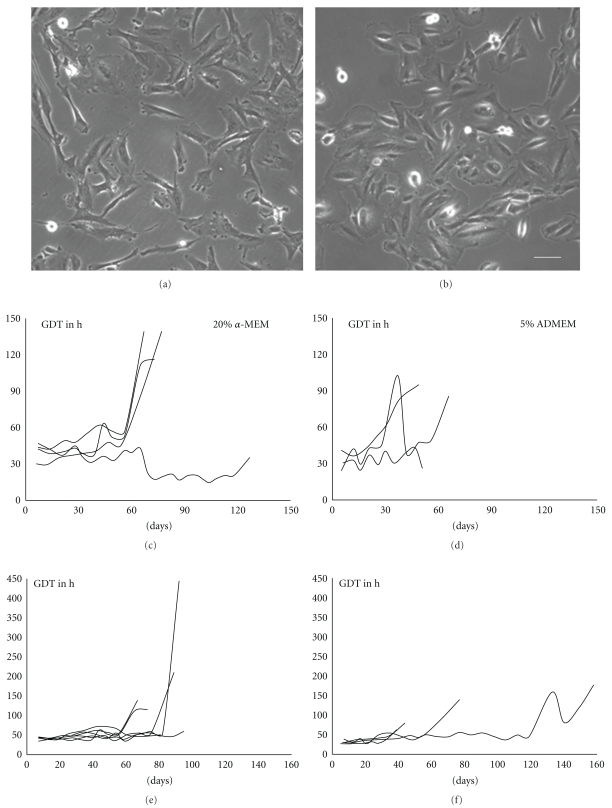
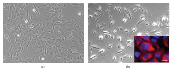
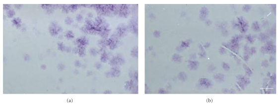
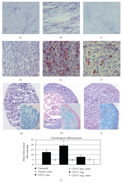
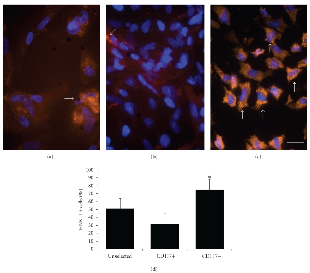
Amniotic fluid (AF) has become an interesting source of fetal stem cells. However, AF contains heterogeneous and multiple, partially differentiated cell types. After isolation from the amniotic fluid, cells were characterized regarding their morphology and growth dynamics. They were sorted by magnetic associated cell sorting using the surface marker CD 117. In order to show stem cell characteristics such as pluripotency and to evaluate a possible therapeutic application of these cells, AF fluid-derived stem cells were differentiated along the adipogenic, osteogenic, and chondrogenic as well as the neuronal lineage under hypoxic conditions. Our findings reveal that magnetic associated cell sorting (MACS) does not markedly influence growth characteristics as demonstrated by the generation doubling time. There was, however, an effect regarding an altered adipogenic, osteogenic, and chondrogenic differentiation capacity in the selected cell fraction. In contrast, in the unselected cell population neuronal differentiation is enhanced.


Figures

Figure 1
Figure 1
Morphology and growth characteristics of AF cells under different culture conditions. (a) AF cells cultivated in the presence of α-MEM supplemented with 10% FBS exhibit an elongated morphology. (b) In the presence of ADMEM with 5% FBS cells revealed a rather epithelioid shape. (c) Cells cultivated in α-MEM (10% FBS) showed a more homogenous generation doubling time (GDT) (d) compared with those cultivated in ADMEM (5% FBS) (n = 5). (e) The GDT of cells derived from amniocenteses is rather homogenous with an average survival time of 60–80 days. (f) Cells derived from amniodrainages reveal somewhat longer survival times than cells derived from amniocenteses and their growth dynamics are inhomogenous (n = 5). Scale bar in a and b = 25 μm.
Figure 2
Figure 2
Morphological analysis of CD117+ and CD117– AF cells. (a) CD117+ cells reveal the typical elongated morphology of mesenchymal stem cells, (b) CD117− AF cells display a rather polymorph appearance of epithelial cells in vitro, inset, CD117− cells can be stained by the epithelial marker cytokeratin (red fluorescence) in a counterstain with the nuclear marker Hoechst dye (blue), scale bar in a and b = 25 μm, and in the inset in b = 15 μm.
Figure 3
Figure 3
CFU assay for the demonstration and comparison of the self-renewing potential of CD117− and CD117+ AF-cells. Plating of very low cell numbers leads to the generation of single colonies (CFU) as demonstrated by the cresyl violet staining. (a) Colony formation after plating of 1000 CD117(−) cells per 100 mm culture dish. (b) Colony formation after plating of 1000 CD117(+) cells per dish. Scale bar = 1 cm.
Figure 4
Figure 4
In vitro differentiation potential of AF cells into the osteogenic (ALP staining), adipogenic, and chondrogenic (Alcian blue staining) lineage. (a) while there was only a weak ALP staining in the CD117− cell fraction, (b) this was markedly stronger in the CD117+ cell population. (c) Unselected AF-cells also reveal a rather weak osteogenic differentiation as demonstrated by a weak ALP staining. (d) While no adipogenic differentiation as demonstrated by the Oil RedO staining could be observed in the CD117– cell fraction, (e) strong positive Oil Red O staining could be observed in the CD117+ fraction, (f) in the unselected population only a minor number of cells showed a Red Oil O positive staining. (g) After chondrogenic induction in CD 117 negative cells there is a diffuse Alcian blue staining detectable. (h) After chondrogenic induction the CD 117 positive cell fraction forms a firm pellet with a strong Alcian blue staining in the cortical zone. The pellets have a hyaline cartilage-like morphology based on characteristic lacunae formation containing round to oval shaped cells which resemble chondrocytes, which occur singly or in nests of two to three cells. (i) Diffuse Alcian blue staining can also be detected in unselected cells. (j) Densitometric analysis of Alcian blue staining in the different experimental groups There is a significant higher staining intensity in the CD117+ cell fraction compared to unselected or CD117− cells (n = 6). Scale bar in (a–i) = 100 μm, in the insets in (g–i) = 50 μm.
Figure 5
Figure 5
Neural marker expression as a proof for the neural differentiation potential. After the neural selection procedure the expression of the neural markers such as (a) α-internexin, (b) βIII tubulin, (c) as well as GFAP can be shown. Secondary antibodies for all primary antibodies are Cy2 conjugated (red fluorescence). Cell nuclei are labelled with the Hoechst dye 33342nuclear stain (blue fluorescence). Scale bar in (a–c) = 10 μm.
Figure 6
Figure 6
HNK-1 expression in AF cells as a confirmation of neuronal differentiation capacity in the CD 117 positive and negative cell fraction and under hypoxic (2% O2) culture conditions. (a) Moderate HNK-1 (orange fluorescence, arrow) expression in unselected cells and (b) a weak expression of the early neuronal marker HNK-1 (arrow). (c) While there is a stronger HNK-1 expression in CD 117 negative cell fraction detectable (arrows), (d) graphical display about the quantification of HNK-1 expression in the unselected, CD117+ and CD117− cell fractions. (∗)significant increase of HNK-1 immunopositive cells compared to control. Secondary antibodies are Cy2 conjugated (red to orange fluorescence. The whole cell population is detected with the Hoechst dye nuclear stain (blue fluorescence) (n = 5, P < .05). Scale bar in (a–f) = 25 μm.
Figure 7
Figure 7
Integration and differentiation of GFP labelled (green fluorescence) predifferentiated CD117− AF cells after grafting into the striatum of adult rats. (a) GFP labelled cells can be found around the injection site in the striatum. (b) one week after grafting GFP expressing cells are still arranged in clusters near the injection site, in a co staining with the astrocyte marker GFAP (Cy2 conjugated secondary antibody, red fluorescence) (c) and (d) typical neural cell morphologies of GFP expressing cells with elongated processes can be detected 2 weeks after grafting in a costaining with the early neuronal marker α-internexin (Cy2,red), (e) a dense network of GFP expressing processes are visible, (f) which show a co-staining with the astrocyte marker GFAP (Cy2, red). (g) Overlay of GFP (green) and GFAP (red) expressing cells. Scale bar in (a) = 20 μm, in (b) = 15 μm and in (c) and (d) = 10 μm.

References

    1. Hemberger M, Yang W, Natale D, et al. Stem cells from fetal membranes—a workshop report. Placenta. 2008;29:S17–S19. - PubMed
    1. de Coppi P, Bartsch G, Jr., Siddiqui MM, et al. Isolation of amniotic stem cell lines with potential for therapy. Nature Biotechnology. 2007;25(1):100–106. - PubMed
    1. Prusa AR, Marton E, Rosner M, Bernaschek G, Hengstschlager M. Oct-4-expressing cells in human amniotic fluid: a new source for stem cell research? Human Reproduction. 2003;18(7):1489–1493. - PubMed
    1. Gosden CM. Amniotic fluid cell types and culture. British Medical Bulletin. 1983;39(4):348–354. - PubMed
    1. Hoehn H, Salk D. Morphological and biochemical heterogeneity of amniotic fluid cells in culture. Methods in Cell Biology. 1982;26:12–34. - PubMed

LinkOut - more resources