. 2011 Mar 17;6(3):e17907.
doi: 10.1371/journal.pone.0017907.

Cooperative nuclear localization sequences lend a novel role to the N-terminal region of MSH6

Affiliations

Cooperative nuclear localization sequences lend a novel role to the N-terminal region of MSH6

Natalie R Gassman et al. PLoS One. .

Abstract

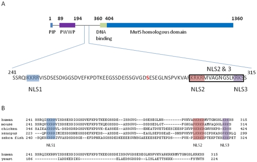
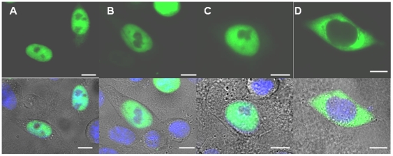
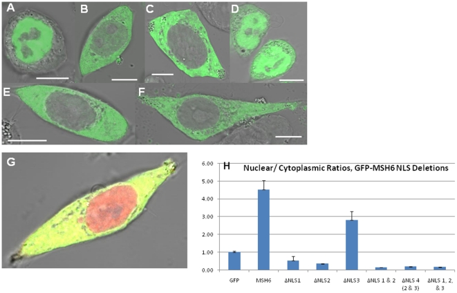
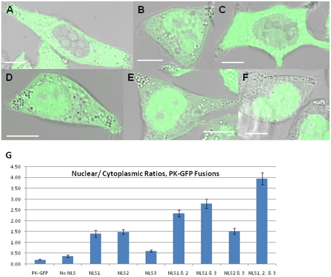
Human mismatch repair proteins MSH2-MSH6 play an essential role in maintaining genetic stability and preventing disease. While protein functions have been extensively studied, the substantial amino-terminal region (NTR*) of MSH6 that is unique to eukaryotic proteins, has mostly evaded functional characterization. We demonstrate that a cluster of three nuclear localization signals (NLS) in the NTR direct nuclear import. Individual NLSs are capable of partially directing cytoplasmic protein into the nucleus; however only cooperative effects between all three NLSs efficiently transport MSH6 into the nucleus. In striking contrast to yeast and previous assumptions on required heterodimerization, human MSH6 does not determine localization of its heterodimeric partner, MSH2. A cancer-derived mutation localized between two of the three NLS significantly decreases nuclear localization of MSH6, suggesting altered protein localization can contribute to carcinogenesis. These results clarify the pending speculations on the functional role of the NTR in human MSH6 and identify a novel, cooperative nuclear localization signal.


Conflict of interest statement

Competing Interests: The authors have declared that no competing interests exist.

Figures

Figure 1
Figure 1. Identification of three nuclear localization sequences in an uncharacterized region of the N-terminal region of MSH6 that are only conserved in higher eukaryotes.
(A) Schematic drawing of human MSH6 detailing its N-terminal region. PCNA interaction motif (PIP) is shown in blue, PWWP domain in purple, and predicted non-specific DNA interaction domain is shown in green. Identified potential nuclear localization sequences are highlighted in blue, red and purple (NLS1, NLS2, and NLS3). Cancer mutation S285I is indicated in bold. (B) Clustal alignment of MSH6 N-terminal regions from different eukaryotic species demonstrating location of the NLSs. Yeast Msh6 contains no comparable region to those found in higher eukaryotes, as shown. The higher eukaryotic species contain highly similar and closely spaced NLSs to those found in human MSH6.
Figure 2
Figure 2. Identification of amino acid stretch in NTR of MSH6 that directs nuclear localization using N-terminal deletion mutants of human GFP-tagged MSH6 in Δmsh6 cells.
Shown are widefield fluorescent images (green) and its overlay with the DIC image to indicate cellular outline. Hoechst staining (blue) indicates the location of the nucleus. All scale bars are 10 µm. (A) GFP-MSH6, full length protein; (B) GFP-MSH6 Δ1–100; (C) GFP-MSH6 Δ1–210; (D) GFP-MSH6 Δ1–399. A clear alteration in nuclear localization is observed upon the truncation of amino acids 211–399.
Figure 3
Figure 3. Deletion of predicted NLS from human GFP-tagged MSH6 in Δmsh6 cells.
Shown are the fluorescent view (green) and its overlay with the DIC image to indicate cellular outline. All scale bars are 10 µm. (A) GFP-MSH6, (B) GFP-MSH6 ΔNLS1, (C) GFP-MSH6 ΔNLS2, (D) GFP-MSH6 ΔNLS3, (E) GFP-MSH6 ΔNLS2/3, (F) GFP-MSH6 ΔNLS1/2/3. (G) Co-transfection with dsRed-MSH2 and GFP-MSH6 Δ1–399, (H) Nuclear to cytoplasmic ratios quantified for the corresponding NLS deletion mutants shown.
Figure 4
Figure 4. Predicted NLSs are validated by their ability to import a cytoplasmic protein, pyruvate kinase, into the nucleus in Δmsh6 cells.
(A) Pyruvate kinase-GFP fusion (PK-GFP); (B) NLS1 PK-GFP; (C) NLS3 PK-GFP; (D) NLS2/3 PK-GFP; (E) NLS 1/3 PK-GFP; (F) (MSH6 NLS1, 2&3)-PK-GFP. (G) Nuclear to cytoplasmic ratios quantified for NLS pyruvate kinase fusions. The no NLS fusion contains only the intervening amino acids between the predicted nuclear localization sequences to validate that this flanking sequence does not accomplish nuclear import in the absence of the predicted NLS.
Figure 5
Figure 5. The full kinetic model for import of MSH6 into the nucleus with 12 species and 14 reactions, is shown diagrammatically.
Each green ball is a molecular species or complex and each yellow oval is a reaction. The arrows into a yellow oval indicate those species are reactants and errors out indicate the species are products. The different species and reaction types are organized spatially for clarity. Each of the four base species, MSH6, and the binders to NLS1, NLS2, and NLS3 (bNLS1, bNLS2, bNLS3, respectively, are shown at the bottom as green circles as species that are reactants for the formation reactions to which they are linked by inward arrows. The formation reactions that lead to complex formation are shown in yellow in the middle, with the complexes formed shown at top as green circles; the complexes formed are linked to the correct formation reactions by outward arrows. The complexes are named appropriately, so that for example, MSH6_b1 is the complex of MSH6 with the binder to NLS1 and is connected via a yellow reaction oval to bNLS1 (the binder to NLS1) and MSH6 in the cytoplasm. The disintegration reactions that implicitly model transport into the nucleus through complex disintegration are shown at top right in yellow, where they connect the appropriate complex to the correct molecular species, leading to MSH6 in the nucleus. For example, MSH6_b1 is connected to MSH6 in the nucleus and bNLS1 (the binder to NLS1) via a disintegration reaction.
Figure 6
Figure 6. A deficiency in MSH6 does not alter the localization of MSH2.
Shown is the localization of endogenous MSH2 in Δmsh6 cells and in Δmsh6 cell transfected with GFP-MSH6. Scale bars are 10 µm. (A) Alexa546 MSH2, (B) Alexa546 MSH2 and GFP-MSH6, (C) Alexa546 MSH2 and GFP-MSH6 ΔNLS 1/2/3.

Similar articles

Cited by

References

    1. Modrich P. Mechanisms in eukaryotic mismatch repair. J Biol Chem. 2006;281:30305–30309. - PMC - PubMed
    1. Kunkel TA, Erie DA. DNA mismatch repair. Annu Rev Biochem. 2005;74:681–710. - PubMed
    1. Hanahan D, Weinberg R. The hallmarks of cancer. Cell. 2000;100:57–70. - PubMed
    1. Harfe B, Jinks-Robertson S. DNA mismatch repair and genetic instability. Annu Rev Genet. 2000;34:359–399. - PubMed
    1. Kolodner R, Marsischky G. Eukaryotic DNA mismatch repair. Curr Opin Genet Dev. 1999;9:89–96. - PubMed

Publication types

MeSH terms

Substances