. 2011 Mar;7(3):e1001317.
doi: 10.1371/journal.ppat.1001317. Epub 2011 Mar 17.

The strain-encoded relationship between PrP replication, stability and processing in neurons is predictive of the incubation period of disease

Affiliations

The strain-encoded relationship between PrP replication, stability and processing in neurons is predictive of the incubation period of disease

Jacob I Ayers et al. PLoS Pathog. 2011 Mar.

Abstract

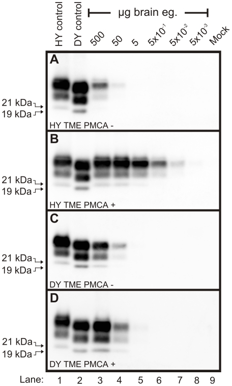
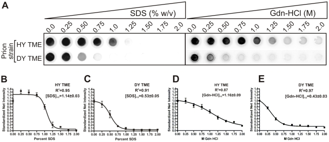
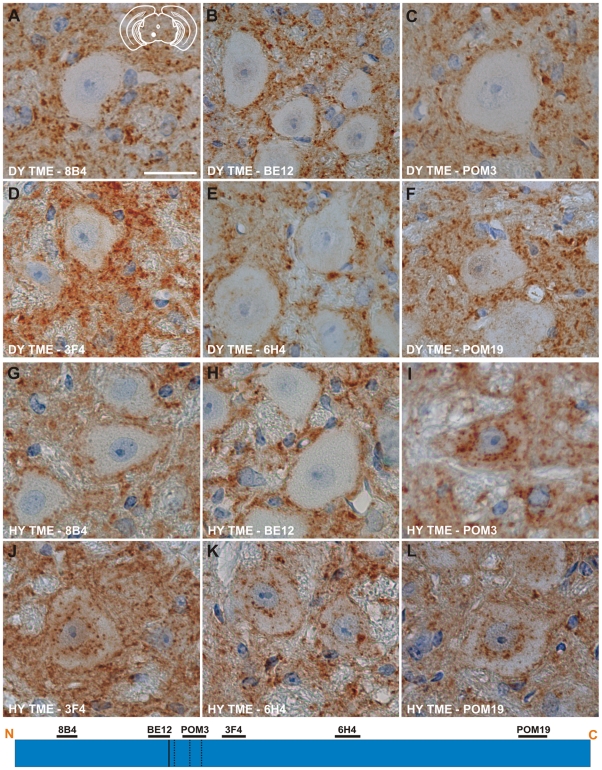
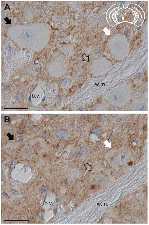
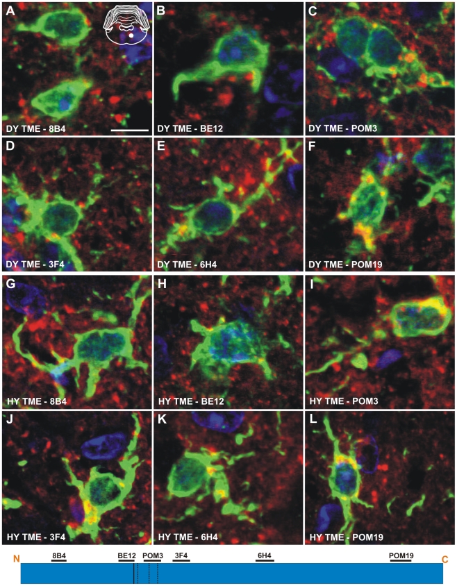
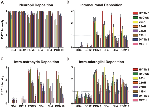
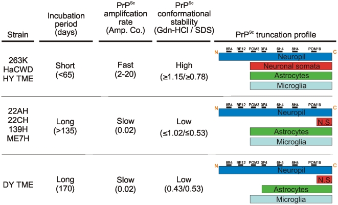
Prion strains are characterized by differences in the outcome of disease, most notably incubation period and neuropathological features. While it is established that the disease specific isoform of the prion protein, PrP(Sc), is an essential component of the infectious agent, the strain-specific relationship between PrP(Sc) properties and the biological features of the resulting disease is not clear. To investigate this relationship, we examined the amplification efficiency and conformational stability of PrP(Sc) from eight hamster-adapted prion strains and compared it to the resulting incubation period of disease and processing of PrP(Sc) in neurons and glia. We found that short incubation period strains were characterized by more efficient PrP(Sc) amplification and higher PrP(Sc) conformational stabilities compared to long incubation period strains. In the CNS, the short incubation period strains were characterized by the accumulation of N-terminally truncated PrP(Sc) in the soma of neurons, astrocytes and microglia in contrast to long incubation period strains where PrP(Sc) did not accumulate to detectable levels in the soma of neurons but was detected in glia similar to short incubation period strains. These results are inconsistent with the hypothesis that a decrease in conformational stability results in a corresponding increase in replication efficiency and suggest that glia mediated neurodegeneration results in longer survival times compared to direct replication of PrP(Sc) in neurons.


Conflict of interest statement

The authors have declared that no competing interests exist.

Figures

Figure 1
Figure 1. The molecular weight and abundance of brain-derived PrPSc of hamster-adapted hamster strains are similar.
A) Western blot analysis and B) quantification (n = 4) of PrPSc from proteinase K digested brain homogenate of hamsters at terminal disease infected with either the HY TME, 263K, HaCWD, 22AH, 22CH, 139H, DY TME or ME7H agents. The migration of the 19 and 21 kDa unglycosylated PrPSc polypeptides is indicated on the left of panel A.
Figure 2
Figure 2. Kinetics of HY and DY PrPSc amplification correspond to differences in incubation period.
Western blot analysis was performed on 10-fold serial dilutions of either HY TME (Panels A and B) or DY TME (Panels C and D) agent infected brain homogenate prior to (Panels A and C) or after one round of PMCA (Panels B and D). The migration of the 19 and 21 kDa unglycosylated PrPSc polypeptides is indicated on the left of each panel. Mock – mock infected negative control reaction.
Figure 3
Figure 3. HY PrPSc is more conformationally stable than DY PrPSc in either Gdn-HCL or SDS.
A) HY or DY TME infected brain homogenate was treated with increasing concentrations of either SDS or Gdn-HCL, digested with PK and the remaining PrPSc was detected using a 96 well immunoassay. The concentration of either SDS or Gdn-HCL required for a 50% reduction in PrPSc is greater for HY TME (panels B and D) compared to DY PrPSc (Panels C and E).
Figure 4
Figure 4. Strain specific differences in the clearance of PrPSc in neurons of hamsters infected with either the DY or HY TME agents.
PrPSc immunohistochemistry was performed on CNS from hamsters infected with either the DY (panels A–F) or the HY TME (panels G–L) agents. PrPSc deposits are detected in the neuropil of hamsters infected with the long incubation period strain DY TME, however, PrPSc was rarely detected in the soma of neurons (panels A–F). In hamsters infected with the short incubation period strain HY TME, PrPSc is detected in the neuropil with all antibodies used (panels G–L). In contrast to the DY TME infected brain, HY PrPSc was detected in the somata of neurons with anti-PrP antibodies whose epitopes are C-terminal to the in vitro PK cleavage site (panels I–L). The yellow region in the schematic insets in panel A depict the location in the brain area that was imaged for every panel. The schematic at the bottom of the figure represents the location of the anti-PrP antibodies and the HY and DY PrPSc PK cleavage sites are depicted as solid and dashed lines, respectively. Scale bar, 50 µm.
Figure 5
Figure 5. Truncation of the N-terminus of HY PrPSc within neurons.
PrPSc immunohistochemistry was performed on serial sections with either an anti-PrP antibody whose epitope is either A) N-terminal (BE12) or B) C-terminal (POM3) to the HY PrPSc PK cleavage site. Arrows indicate the same neurons in panels A and B. The yellow region in the schematic insets depict the location in the brain area that was imaged in each panel. Abbreviations: b.v., blood vessels; w.m., white matter. Scale bar, 50 µm.
Figure 6
Figure 6. Strain-specific truncation of PrPSc in astrocytes of hamsters infected with either the DY or HY TME agents.
Dual immunofluorescence was performed on brains of DY TME (panels A–F) or HY TME (panels G–L) infected animals using antibodies directed against PrP (red fluorescence) and GFAP (green fluorescence). Dual PrP/GFAP immunofluorescence was performed on the reticular formation from DY TME (A–F) or HY TME (G–L) agent infected hamsters at the clinical stage of disease. The solid white circle located in the schematic inset is the location of the photographed images within the reticular formation. The schematic at the bottom of the figure represents the location of the anti-PrP antibodies and the HY and DY PrPSc PK cleavage sites are depicted as solid and dashed lines, respectively. The HY and DY PrPSc PK cleavage sites are also depicted as the solid and dashed lines respectively. Scale bar, 10 µm.
Figure 7
Figure 7. Identical processing of PrPSc in microglia of hamsters infected with either the DY or HY TME agents.
Double immunofluorescence was performed using antibodies directed against PrP (red fluorescence) and Iba-1 (green fluorescence). (A–F) Immunofluorescence in the reticular formation of DY TME infected hamsters at the clinical stage of disease. (G–L) Immunofluorescence in the reticular formation of HY TME infected hamsters at the clinical stage of disease. The solid white circle located in the schematic inset is the location of the brain area that was imaged in each panel. The schematic at the bottom of the figure represents the location of the anti-PrP antibodies and the HY and DY PrPSc PK cleavage sites are depicted as solid and dashed lines, respectively. Scale bar, 10 µm.
Figure 8
Figure 8. Strain-specific processing of PrPSc in neurons and glia.
The intensity of PrPSc immunoreactivity for each hamster-adapted prion strain was scored following immunohistochemistry using either 8B4, BE12, POM3, 3F4, 6H4 or POM19 anti-PrP antibodies. PrPSc deposition was scored in the neuropil (A), within neurons (B), astrocytes (C), and microglia (D). Bars represent mean PrPSc intensity values and lines represent standard error.
Figure 9
Figure 9. The relationship between PrPSc replication, stability and deposition in neurons is predictive of the incubation period of disease.
The prion strains were grouped according to commonalities in incubation period, PrPSc amplification rate, PrPSc conformational stability and PrPSc truncation profile in neurons and glia. N.S. – neuronal somata.

Similar articles

Cited by

References

    1. Castilla J, Saa P, Hetz C, Soto C. In vitro generation of infectious scrapie prions. Cell. 2005;121:195–206. - PubMed
    1. Deleault NR, Harris BT, Rees JR, Supattapone S. Formation of native prions from minimal components in vitro. Proc Natl Acad Sci U S A. 2007;104:9741–9746. - PMC - PubMed
    1. Prusiner SB. Novel proteinaceous infectious particles cause scrapie. Science. 1982;216:136–144. - PubMed
    1. Wang F, Wang X, Yuan CG, Ma J. Generating a prion with bacterially expressed recombinant prion protein. Science. 2010;327:1132–1135. - PMC - PubMed
    1. Makarava N, Kovacs GG, Bocharova O, Savtchenko R, Alexeeva I, et al. Recombinant prion protein induces a new transmissible prion disease in wild-type animals. Acta Neuropathol. 2010;119:177–187. - PMC - PubMed

Publication types

MeSH terms