

Multiple cancer/testis antigens are preferentially expressed in hormone-receptor negative and high-grade breast cancers
- PMID: 21437249
- PMCID: PMC3060908
- DOI: 10.1371/journal.pone.0017876
Multiple cancer/testis antigens are preferentially expressed in hormone-receptor negative and high-grade breast cancers
Abstract
Background: Cancer/testis (CT) antigens are protein antigens normally expressed only in germ cells of testis, and yet are expressed in a proportion of a wide variety of human cancers. CT antigens can elicit spontaneous immune responses in cancer patients with CT-positive cancers, and CT antigen-based therapeutic cancer vaccine trials are ongoing for "CT-rich" tumors. Although some previous studies found breast cancer to be "CT-poor", our recent analysis identified increased CT mRNA transcripts in the ER-negative subset of breast cancer.
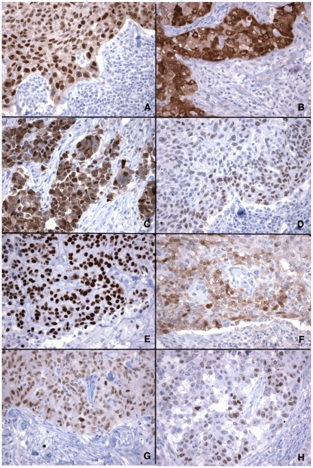
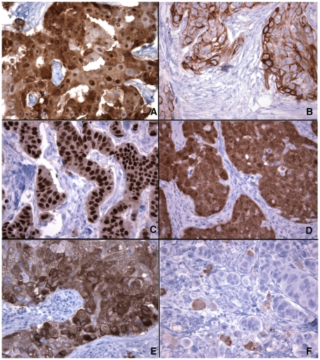
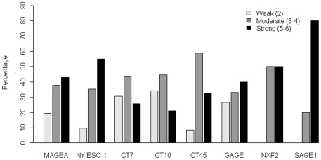
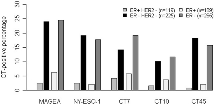
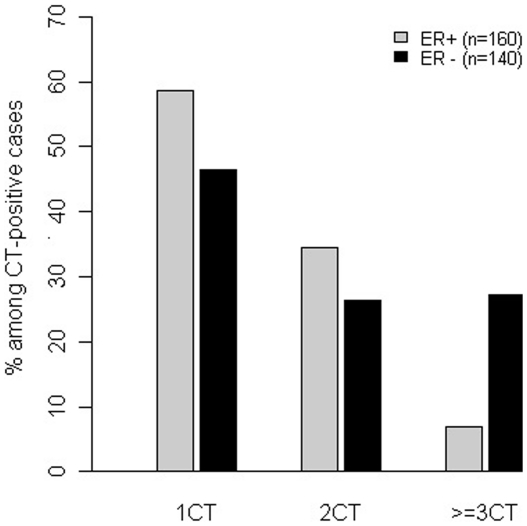
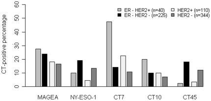
Methodology/principal findings: In this study, we performed a comprehensive immunohistochemical study to investigate the protein expression of eight CT genes in 454 invasive ductal carcinomas, including 225 ER/PR/HER2-negative (triple-negative) carcinomas. We found significantly more frequent expression of all eight CT antigens in ER-negative cancers, and five of them--MAGEA, CT7, NY-ESO-1, CT10 and CT45, were expressed in 12-24% of ER-negative cancers, versus 2-6% of ER-positive cancers (p<0.001 to 0.003). In comparison, GAGE, SAGE1 and NXF2 were only expressed in 3-5% of ER-negative and 0-2% of ER-positive cancers. ER-negative cancers were also more likely to simultaneously co-express multiple CT antigens, with 27% (34/125) of ER-negative, CT-positive tumors expressing three or more CT antigens. HER2 status had no consistent effect on CT expression, and triple-negative carcinomas showed similar frequencies of MAGEA and NY-ESO-1 expression as ER-negative/HER2-positive carcinomas. More frequent CT expression was also found in tumors with higher nuclear grade (p<0.001 to p = 0.01) and larger in size (>2 cm).
Conclusions/significance: CT antigens are preferentially expressed in hormone receptor-negative and high-grade breast cancer. Considering the limited treatment options for ER/PR/HER2 triple-negative breast cancer, the potential of CT-based immunotherapy should be explored.
Conflict of interest statement
Figures
References
-
- Boon T, Coulie PG, Van den Eynde B. Tumor antigens recognized by T cells. Immunol Today. 1997;18:267–268. - PubMed
-
- Scanlan MJ, Gure AO, Jungbluth AA, Old LJ, Chen YT. Cancer/testis antigens: an expanding family of targets for cancer immunotherapy. Immunol Rev. 2002;188:22–32. - PubMed
-
- Simpson AJ, Caballero OL, Jungbluth A, Chen YT, Old LJ. Cancer/testis antigens, gametogenesis and cancer. Nat Rev Cancer. 2005;5:615–625. - PubMed
-
- van der Bruggen P, Traversari C, Chomez P, Lurquin C, De Plaen E, et al. A gene encoding an antigen recognized by cytolytic T lymphocytes on a human melanoma. Science. 1991;254:1643–1647. - PubMed
Publication types
MeSH terms
Substances
LinkOut - more resources
Full Text Sources
Other Literature Sources
Medical
Research Materials
Miscellaneous
