. 2011 Jun;69(6):954-62.
doi: 10.1002/ana.22348. Epub 2011 Mar 17.

Arteriovenous malformation in the adult mouse brain resembling the human disease

Affiliations

Arteriovenous malformation in the adult mouse brain resembling the human disease

Espen J Walker et al. Ann Neurol. 2011 Jun.

Abstract

Objective: Brain arteriovenous malformations (bAVMs) are an important cause of hemorrhagic stroke. The underlying mechanisms are not clear. No animal model for adult bAVM is available for mechanistic exploration. Patients with hereditary hemorrhagic telangiectasia type 2 (HHT2) with activin receptor-like kinase 1 (ALK1; ACVRL1) mutations have a higher incidence of bAVM than the general population. We tested the hypothesis that vascular endothelial growth factor (VEGF) stimulation with regional homozygous deletion of Alk1 induces severe dysplasia in the adult mouse brain, akin to human bAVM.

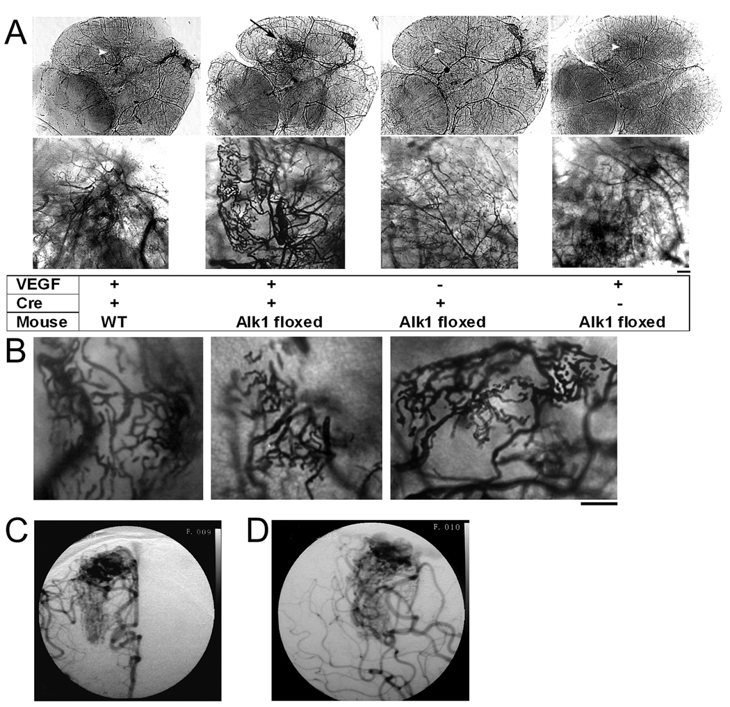
Methods: Alk1(2f/2f) (exons 4-6 flanked by loxP sites) and wild-type (WT) mice (8-10 weeks old) were injected with adenoviral vector expressing Cre recombinase (Ad-Cre; 2 × 10(7) plaque forming units [PFU]) and adeno-associated viral vectors expressing VEGF (AAV-VEGF; 2 × 10(9) genome copies) into the basal ganglia. At 8 weeks, blood vessels were analyzed.

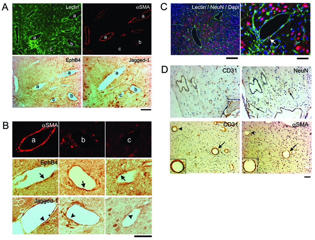
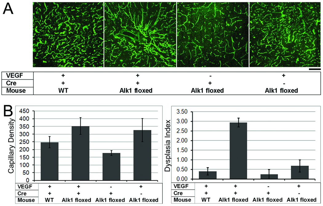
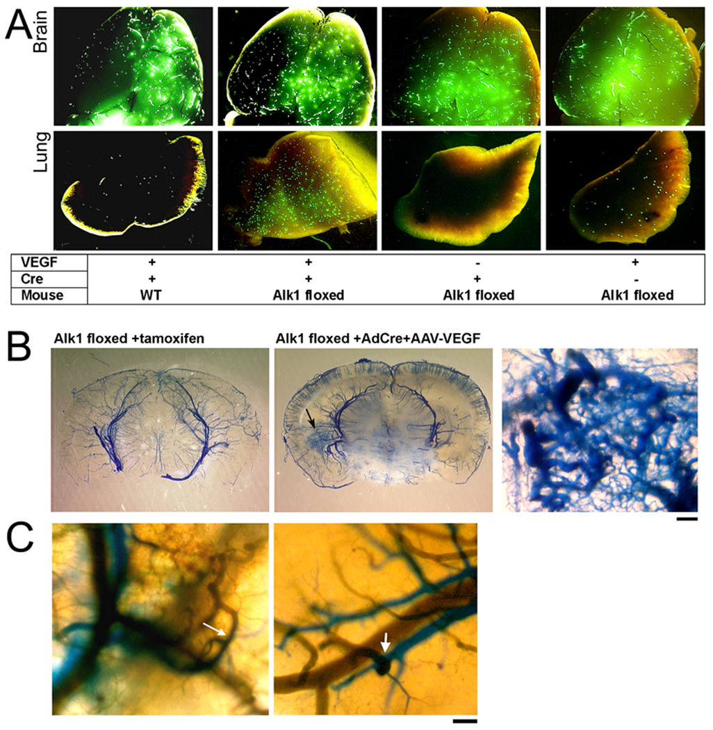
Results: Gross vascular irregularities were seen in Alk1(2f/2f) mouse brain injected with Ad-Cre and AAV-VEGF. The vessels were markedly enlarged with abnormal patterning resembling aspects of the human bAVM phenotype, displayed altered expression of the arterial and venous markers (EphB4 and Jagged-1), and showed evidence of arteriovenous shunting. Vascular irregularities were not seen in similarly treated WT mice.

Interpretation: Our data indicate that postnatal, adult formation of the human disease, bAVM, is possible, and that both genetic mutation and angiogenic stimulation are necessary for lesion development. Our work not only provides a testable adult mouse bAVM model for the first time, but also suggests that specific medical therapy can be developed to slow bAVM growth and potentially stabilize the rupture-prone abnormal vasculature.


Figures

Figure 1
Figure 1. Vessel casting showing that VEGF stimulation induced distinct cerebrovascular abnormalities at the Alk1-deleted region
(A) Large tangled vessels resembling a bAVM were detected at the injection site of Ad-Cre and AAV-VEGF in the brain of Alk1-floxed mice (black arrow). Alk1 deletion (Ad-Cre injection) without VEGF had no effect on the cerebrovascular structure. Overexpression of VEGF in the brain without Alk1 deletion (WT mice or Alk1 mice injected with control adenoviral vector) induced normal angiogenesis. The bottom images show the enlarged angiogenic foci of the images on top. Scale bar = 100 µm. Injection sites are indicated by white arrow heads. (B) Abnormal vasculature from 3 different Alk1-floxed mice injected with Ad-Cre and AAV-VEGF. Scale bar = 100 µm. (C) & (D) Right internal carotid artery anterior-posterior (C) and lateral (D) projections of an angiogram from an 18-year-old male who underwent microsurgical bAVM resection. The bAVM, supplied by the middle and anterior cerebral arteries, has a diffuse angioarchitecture, similar to the phenotype in the mouse model.
Figure 2
Figure 2. Dysplastic vessels have irregular smooth muscle coverage and express inconsistent arterial and venous markers
(A) A group of dilated irregular vessels labeled with lectin (green). Some of these vessels have smooth muscle coverage (αSMA, red), some do not. Arterial (Jagged-1) and venous (EphB4) markers were expressed by the endothelial cells of vessels with or without smooth muscle. Scale Bar: 50 µm. (B) Enlarged image of the vessel (a), (b), (c) indicated in (A). Vessel (a) is covered with a complete layer of smooth muscle and (b) has a few αSMA positive cells. Their endothelial cells express EphB4 and Jagged-1 (arrows and arrowheads). Vessel (c) has almost no αSMA positive cells. Most of its endothelial cells express EphB4, a few express Jagged-1. Scale Bar: 50 µm. (C) Neuronal tissue is present between enlarged irregular vessels. Arrow indicates NeuN positive cell on the dysplastic vessel wall. Scale Bars: 200 µm (left) and 50 µm (right). Corresponding colors for Lectin, NeuN, and Dapi are green, red, and blue, respectively. (D) Paraffin sections of surgical specimen from patient shown in Figure 1, stained with antibodies specific to CD31, αSMA or NeuN. Top panel shows NeuN positive cells (arrows) detected between the dysplastic vessels. Lower panel shows enlarged vessels with (arrows) or without (arrowheads) smooth muscle layers. Inserts in the lower panel show enlarged images of the vessels indicated by arrows with endothelial cells stained by CD31 antibody and a thin layer of smooth muscle (αSMA) surrounding the endothelial layer. Scale Bar: 50 µm.
Figure 3
Figure 3. VEGF induced focal angiogenesis in the normal brain and dysplastic vessel formation in the Alk1-deleted brain
(A) Representative images show lectin-perfused vessels in the brain. Capillary density is high in all AAV-VEGF-treated groups as compared to the AAV-LacZ control group. Dilated irregular vessels were only observed in the brain with Alk1 deletion and VEGF stimulation. Scale Bar=50 µm. (B) Bar graphs show capillary density (left) and dysplasia index (right). Capillary density increased in all groups with VEGF stimulation as compared to the AAV-lacZ control group (p=0.07). Dysplasia index increased significantly in Alk1-floxed mice treated with Ad-Cre and AAV-VEGF as compared to all other groups (p<0.01).
Figure 4
Figure 4. Arteriovenous (A–V) shunting was detected in the brain with regional Alk1 deletion and VEGF stimulation
(A) Brain and lung samples collected from animals perfused with 20 µm fluorescent beads through the carotid artery. Beads were detected in all brain samples, but only Alk1-floxed mice injected with Ad-Cre and AAV-VEGF had a significant amount of beads in the lung. A few beads were detected in the lungs of control mice injected with AAV-VEGF. (B) Latex-perfused brain. Latex (blue) presented only in cerebral arteries of Alk12f/2f/ROSA26(+/creER) mice treated with tamoxifen that had Alk1 gene deleted globally (left). Alk1-floxed mice that received intracerebral injection of Ad-Cre and AAV-VEGF had an increase of vascular density at the injection site (arrow, middle image). At higher magnification (right), vessels are enlarged and tortuous with a fistula between arteries and veins. Scale Bar = 100 µm. (C) Two color latex perfusion shows direct connections of cerebral arteries (blue) and veins (green) (arrows). Scale Bar = 100 µm.

References

    1. Kim H, Marchuk DA, Pawlikowska L, et al. Genetic considerations relevant to intracranial hemorrhage and brain arteriovenous malformations. Acta Neurochir Suppl. 2008;105:199–206. - PMC - PubMed
    1. Fleetwood IG, Steinberg GK. Arteriovenous malformations. Lancet. 2002;359:863–873. - PubMed
    1. Mullan S, Mojtahedi S, Johnson DL, Macdonald RL. Embryological basis of some aspects of cerebral vascular fistulas and malformations. J Neurosurg. 1996;85:1–8. - PubMed
    1. Kim H, Sidney S, McCulloch CE, et al. Racial/ethnic differences in longitudinal risk of intracranial hemorrhage in brain arteriovenous malformation patients. Stroke. 2007;38:2430–2437. - PubMed
    1. Kim H, Pawlikowska L, Young WL. Genetics and vascular biology of brain vascular malformations. In: Mohr JP, Wolf PA, Grotta JC, et al., editors. Stroke: Pathophysiology, Diagnosis, and Management. 5th ed. Philadelphia: Churchill Livingstone Elsevier; 2010.

Publication types

MeSH terms