Different structural correlates for verbal memory impairment in temporal lobe epilepsy with and without mesial temporal lobe sclerosis
- PMID: 21438080
- PMCID: PMC3259857
- DOI: 10.1002/hbm.21226
Different structural correlates for verbal memory impairment in temporal lobe epilepsy with and without mesial temporal lobe sclerosis
Abstract
Objectives: Memory impairment is one of the most prominent cognitive deficits in temporal lobe epilepsy (TLE). The overall goal of this study was to explore the contribution of cortical and hippocampal (subfield) damage to impairment of auditory immediate recall (AIMrecall), auditory delayed recall (ADMrecall), and auditory delayed recognition (ADMrecog) of the Wechsler Memory Scale III (WMS-III) in TLE with (TLE-MTS) and without hippocampal sclerosis (TLE-no). It was hypothesized that volume loss in different subfields determines memory impairment in TLE-MTS and temporal neocortical thinning in TLE-no.
Methods: T1 whole brain and T2-weighted hippocampal magnetic resonance imaging and WMS-III were acquired in 22 controls, 18 TLE-MTS, and 25 TLE-no. Hippocampal subfields were determined on the T2 image. Free surfer was used to obtain cortical thickness averages of temporal, frontal, and parietal cortical regions of interest (ROI). MANOVA and stepwise regression analysis were used to identify hippocampal subfields and cortical ROI significantly contributing to AIMrecall, ADMrecall, and ADMrecog.
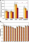
Results: In TLE-MTS, AIMrecall was associated with cornu ammonis 3 (CA3) and dentate (CA3&DG) and pars opercularis, ADMrecall with CA1 and pars triangularis, and ADMrecog with CA1. In TLE-no, AIMrecall was associated with CA3&DG and fusiform gyrus (FUSI), and ADMrecall and ADMrecog were associated with FUSI.
Conclusion: The study provided the evidence for different structural correlates of the verbal memory impairment in TLE-MTS and TLE-no. In TLE-MTS, the memory impairment was mainly associated by subfield-specific hippocampal and inferior frontal cortical damage. In TLE-no, the impairment was associated by mesial-temporal cortical and to a lesser degree hippocampal damage.
Copyright © 2011 Wiley Periodicals, Inc.
Figures


References
-
- Acsady L, Kali S ( 2007): Models, structure, function: The transformation of cortical signals in the dentate gyrus. Prog Brain Res 163: 577–599. - PubMed
-
- Aggleton J, Brown MW ( 2006): Interleaving brain system for episodic and recognition memory. Trends Cogn Sci 10: 455–463. - PubMed
-
- Alessio A, Damasceno BP, Camargo CHP, Kobayashi E, Guerreiro CAM, Cendes F ( 2004): Differences in memory performance and other clinical characteristics in patients with mesial temporal lobe epilepsy with and without hippocampal atrophy. Epilepsy Behav 5: 22–27. - PubMed
-
- Anderson KL, Rajagovindan R, Ghacibeh GA, Meador KJ, Ding M ( 2010): Theta oscillations mediate interaction between prefrontal cortex and medial temporal lobe in human memory. Cereb Cortex 20: 1604–1612. - PubMed
-
- Bernhardt BC, Worsley KJ, Besson P, Concha L, Lerch JP, Evans AC, Bernasconi N ( 2008): Mapping limbic network organization in temporal epilepsy using morphometric correlations: Insights on the relation between mesiotemporal connectivity and cortical atrophy. Neuroimage 42: 515–524. - PubMed
Publication types
MeSH terms
Grants and funding
LinkOut - more resources
Full Text Sources
Medical
Miscellaneous

