. 2011 Mar 26:9:28.
doi: 10.1186/1741-7015-9-28.

Nuclear envelope structural defects cause chromosomal numerical instability and aneuploidy in ovarian cancer

Affiliations

Nuclear envelope structural defects cause chromosomal numerical instability and aneuploidy in ovarian cancer

Callinice D Capo-chichi et al. BMC Med. .

Abstract

Background: Despite our substantial understanding of molecular mechanisms and gene mutations involved in cancer, the technical approaches for diagnosis and prognosis of cancer are limited. In routine clinical diagnosis of cancer, the procedure is very basic: nuclear morphology is used as a common assessment of the degree of malignancy, and hence acts as a prognostic and predictive indicator of the disease. Furthermore, though the atypical nuclear morphology of cancer cells is believed to be a consequence of oncogenic signaling, the molecular basis remains unclear. Another common characteristic of human cancer is aneuploidy, but the causes and its role in carcinogenesis are not well established.

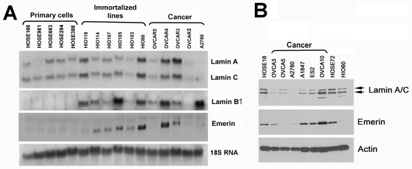
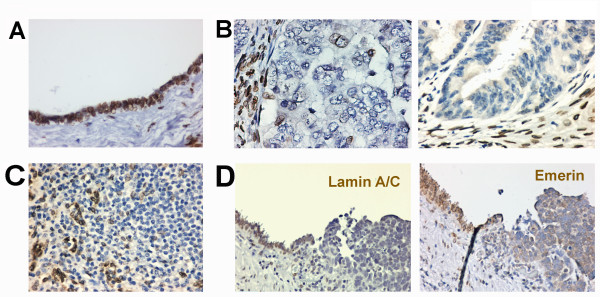
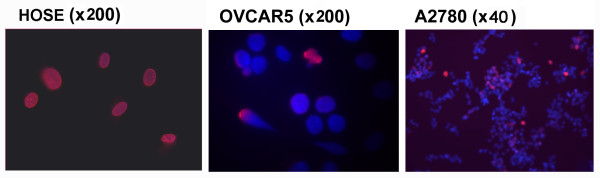
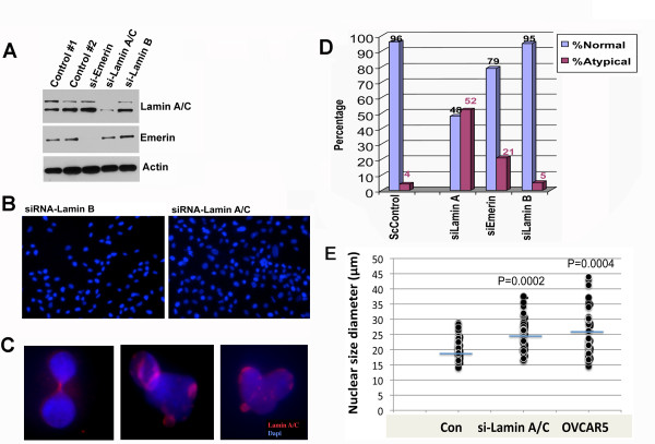
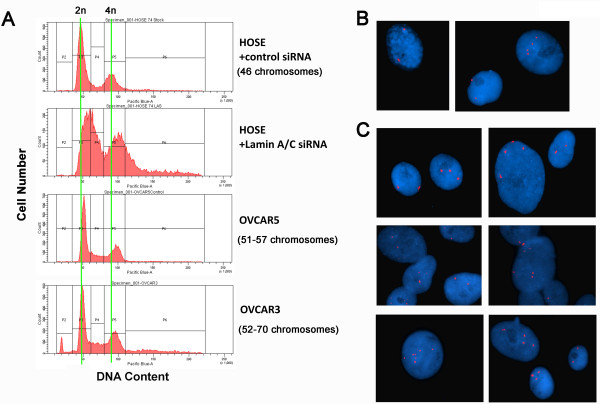
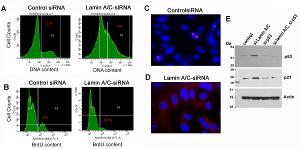
Methods: We investigated the expression of the nuclear envelope proteins lamin A/C in ovarian cancer by immunohistochemistry and studied the consequence of lamin A/C suppression using siRNA in primary human ovarian surface epithelial cells in culture. We used immunofluorescence microscopy to analyze nuclear morphology, flow cytometry to analyze cellular DNA content, and fluorescence in situ hybridization to examine cell ploidy of the lamin A/C-suppressed cells.

Results: We found that nuclear lamina proteins lamin A/C are often absent (47%) in ovarian cancer cells and tissues. Even in lamin A/C-positive ovarian cancer, the expression is heterogeneous within the population of tumor cells. In most cancer cell lines, a significant fraction of the lamin A/C-negative population was observed to intermix with the lamin A/C-positive cells. Down regulation of lamin A/C in non-cancerous primary ovarian surface epithelial cells led to morphological deformation and development of aneuploidy. The aneuploid cells became growth retarded due to a p53-dependent induction of the cell cycle inhibitor p21.

Conclusions: We conclude that the loss of nuclear envelope structural proteins, such as lamin A/C, may underlie two of the hallmarks of cancer--aberrations in nuclear morphology and aneuploidy.


Figures

Figure 1
Figure 1
Expression of nuclear envelope proteins in ovarian epithelial and cancer cell lines. Primary non-cancer cells and cancer cell lines were analyzed for mRNA and protein expression of nuclear envelope proteins lamin A/C, lamin B1, and emerin. (A) Total RNA was isolated and analyzed by Northern blot of primary human ovarian surface epithelial cells (HOSE166, HOSE981, HOSE663, HOSE294, and HOSE388), immortalized lines (HIO118, HIO114, HIO107, HIO105, HIO80), and ovarian cancer cells (OVCAR8, OVCAR4, OVCAR3, OVCAR5, and A2780) for the expression of lamin A/C, lamin B1, and emerin. (B) Total cell lysates from two lines of primary ovarian surface epithelial cells (HOSE18, HOSE72), an immortalized line (HIO60), and six ovarian cancer lines (OVCAR3, OVCAR5, OVCAR10, A2780, A1847, and ES2), were prepared from cultures for Western blot analysis.
Figure 2
Figure 2
Loss of lamin A/C expression in ovarian cancers. Ovarian normal and tumor tissues were analyzed by immunostaining for lamin A/C. (A) A representative example shows that lamin A/C is strongly stained around the nucleus of normal ovarian surface epithelial cells. The nuclei are generally smooth and oval-shaped in all normal ovarian surface epithelial cells. (B) Two examples show loss of lamin A/C expression in ovarian carcinomas. Some stromal cells show strong lamin A/C staining around the nucleus, serving as an internal positive control. The nuclear morphology of tumor cells is heterogeneous, and some cells with large and deformed nuclei are present. (C) A representative example of ovarian carcinomas showing heterogeneous expression of lamin A/C among the tumor cells. (D) An example of ovarian carcinomas contiguous with the benign ovarian surface epithelium. Lamin A/C is lost in the transformed cells. In an adjacent section, the loss of lamin A/C correlates with the distribution of emerin to the cytoplasm of tumor cells.
Figure 3
Figure 3
Heterogeneous lamin A/C expression in ovarian cancer cells. Lamin A/C expression in ovarian normal and cancer epithelial cells was analyzed by immunofluorescence microscopy for DAPI (blue) and lamin A/C (red). Primary ovarian surface epithelial (HOSE) cells exhibit a uniform staining pattern for lamin A/C. All ovarian cancer cell lines show heterogeneous staining of lamin A/C within the cell population, as shown in examples, the OVCAR5 and A2780 ovarian cancer cells.
Figure 4
Figure 4
Suppression of lamin A/C expression in ovarian surface epithelial cells leads to a deformed nuclear shape. Primary HOSE cells were treated with siRNA to lamin A/C for three days and were analyzed for the effect on nuclear size and morphology. (A) Western blot shows the specific suppression of protein by siRNA to emerin, lamin A/C, and lamin B1 in primary HOSE cells. (B) A low magnification image (× 200) shows the presence of a high percentage of cells with large and deformed nuclear morphology following suppression of lamin A/C by siRNA in primary HOSE cells for three days. Suppression of lamin B had no significant effect on nuclear morphology. (C) Examples shown at high magnification (× 1,000) of individual deformed nuclei with reduced and patchy lamin A/C staining. (D) The nuclear morphological abnormality was scored by computer-assisted image analysis. About 100 nuclei in each group treated by siRNA were analyzed and the percentage of normal and atypical nuclear morphology is shown. ScControl: scrambled controls. (E) The nuclear size/diameter was scored by computer-assisted image analysis (AxioVision software 4) following shRNA treatment for 3 days. Nuclear sizes were plotted following computer-assisted imaging analysis of 50 nuclei randomly selected from each group, compared to OVCAR5 ovarian cancer cells.
Figure 5
Figure 5
Lamin A/C suppression in primary ovarian epithelial cells results in increased nuclear size, aneuploidy, and polyploidy. Primary HOSE cells were transfected with control or shRNA to suppress lamin A/C and were subjected to analysis for DNA content and ploidy. (A) The shRNA-treated and control HOSE cells were sorted by flow cytometry to compare DNA content. Two aneuploid ovarian cancer cell lines, OVCAR5 (51 to 57 chromosomes per cell) and OVCAR3 (52 to 70 chromosomes per cell) were analyzed for comparison. A subtle increase in DNA content was observed in lamin A/C-suppressed HOSE cells. (B) In situ hybridization with X chromosome centromere probe of control HOSE cells treated with scrambled shRNA. Two representative fields are shown. (C) In situ hybridization with X centromere probe of lamin A/C-suppressed HOSE cells. Six representative fields are shown for the presence of 3, 4, and other multiple X-chromosome-containing nuclei.
Figure 6
Figure 6
Lamin A/C suppression in primary ovarian epithelial cells results in growth retardation. Primary HOSE cells were transfected with control or shRNA to suppress lamin A/C expression. The cells were analyzed for DNA content and bromodeoxyuridine (BrdU) incorporation simultaneously by flow cytometry. (A) Primary HOSE cells were analyzed by flow cytometry for DNA content following treatment with control or lamin A/C shRNA for three days. Lamin A/C-suppressed cells have > 2n DNA in 75% of cells, compared to 23% of control cells. (B) The same cell populations were incubated with BrdU for one hour in culture, and then fixed and stained with FITC-anti-BrdU prior to flow cytometry to determine BrdU incorporation as an indicator of proliferation. (C) The control cells show a high percentage of mitotic nuclei as shown by immunofluorescence microscopy of beta-tubulin staining of the mitotic spindles. (D) In comparison, beta-tubulin-immunostained mitotic spindles are rare in lamin A/C-suppressed cells. (E) The HOSE cells were treated with siRNA-p53 and/or siRNA-lamin A/C for three days and then analyzed for p53 and p21 by Western blot.

References

    1. Zink D, Fischer AH, Nickerson JA. Nuclear structure in cancer cells. Nat Rev Cancer. 2004;4:677–687. doi: 10.1038/nrc1430. - DOI - PubMed
    1. Pienta KJ, Coffey DS. Correlation of nuclear morphometry with progression of breast cancer. Cancer. 1991;68:2012–2016. doi: 10.1002/1097-0142(19911101)68:9<2012::AID-CNCR2820680928>3.0.CO;2-C. - DOI - PubMed
    1. Papanicolaou GN. A new procedure for staining vaginal smears. Science. 1942;95:438–439. doi: 10.1126/science.95.2469.438. - DOI - PubMed
    1. Hsu CY, Kurman RJ, Vang R, Wang TL, Baak J, Shih IeM. Nuclear size distinguishes low- from high-grade ovarian serous carcinoma and predicts outcome. Hum Pathol. 2005;36:1049–1054. doi: 10.1016/j.humpath.2005.07.014. - DOI - PubMed
    1. Dehari R, Kurman RJ, Logani S, Shih IeM. The development of high-grade serous carcinoma from atypical proliferative (borderline) serous tumors and low-grade micropapillary serous carcinoma: a morphologic and molecular genetic analysis. Am J Surg Pathol. 2007;31:1007–1012. doi: 10.1097/PAS.0b013e31802cbbe9. - DOI - PubMed

Publication types